Dental Corner: Properly equip your dental suite
The key to providing high-quality oral care is having the knowledge and skills to recommend and deliver appropriate treatment, but without the proper equipment, your ability to perform basic dental procedures, such as periodontal prophylaxis and dental extraction, is compromised.
You have made a commitment to yourself and your patients to upgrade your dental services. After all, periodontal disease is the most common disease in dogs.1,2 The key to providing high-quality oral care is having the knowledge and skills to recommend and deliver appropriate treatment, but without the proper equipment, your ability to perform basic dental procedures, such as periodontal prophylaxis and dental extraction, is compromised.
The American Animal Hospital Association (AAHA) recently established dentistry guidelines for accredited hospitals.3 The guidelines state that all hospitals should have equipment to expose and develop intraoral dental radiographs, a high and low-speed air and water delivery system as well as handpieces, dental burs, and a powered scaler. Dental instruments considered necessary by the AAHA guidelines include hand scalers and curettes, a periodontal probe and explorer, and the various instruments needed for dental extractions. In addition, the guidelines state that the instruments should be sterilized between uses and that patients undergoing dental cleanings or extractions should receive inhalation anesthesia with a cuffed endotracheal tube.3
Properly equipping your practice for dentistry is now the accepted standard of care. In this article, we list the equipment you need to perform periodontal prophylaxis and dental extraction (Table 1). This list is not intended to recommend or endorse any particular brand or manufacturer—for specific recommendations, contact your colleagues who have already purchased equipment, or contact a diplomate of the American Veterinary Dental College.


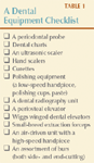
Table 1: A Dental Equipment Checklist
EQUIPMENT FOR GENERAL PERIODONTAL PROPHYLAXIS
These basic instruments are vital to a properly performed periodontal prophylaxis, keeping dental problems at bay.
Periodontal probe and dental explorer
When equipping your dental operatory, the instrument to put at the top of your list is a periodontal probe. Not only is it likely to be the least expensive dental instrument, but it's also the most sensitive clinical tool for detecting periodontal attachment loss.4 The periodontal probe is a blunt-tipped instrument with graduated markings (different probes have different graduations). This probe measures the extent of periodontal attachment loss (Figure 1). It is gently inserted between the free gingiva and tooth to measure pocket depth and gingival recession. Many probes also have a dental explorer at the opposite end of the handle. This sharp, curved instrument, which looks like a shepherd's hook, is valuable in detecting irregularities in teeth such as fractures, caries, and feline resorptive lesions. It can also be used to detect tooth mobility that may be a sign of periodontal disease, tooth root fracture, or neoplasia.


Figure 1. This Marquis color-coded periodontal probe (right), marked in 3-mm increments, is used to measure periodontal attachment loss. The shepherd's hook explorer at the opposite end (left) is used for discovering tooth defects such as cavities and fractures.
Dental chart
Gone are the days when a dental chart consisted of the word Dental beside a check box. Every patient undergoing a dental procedure should have a complete oral examination, and the results of this examination must be entered into the medical record on a dental chart. Additionally, it is important to record the details of any treatments performed, including a description of which teeth were treated, which treatments were performed, and what materials were used. The little stickers with the picture of the teeth on them are better than nothing, but a full-page chart allows for neat notes without crowding. Samples of canine and feline dental charts are available for download at www.dacross.com or, for members of the Veterinary Information Network, at www.vin.com.
Ultrasonic scaler
Ultrasonic scalers are efficient tools for plaque and calculus removal. The units can be powered by stacked magnetostrictive leaves, a magnetostrictive rod, or a piezoelectric signal. The result is tip movement either in an elliptical or curvilinear fashion that produces ultrasonic vibrations (Figure 2). Because heat is generated during the process, a water spray is used to cool the tip. The effects of the ultrasonic sound waves on the water also kill oral bacteria through a process called cavitation.5 To avoid thermal necrosis or inflammation of the sensitive pulp, the tip should only contact each tooth for a few seconds at a time.


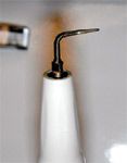
Figure 2. A standard prophylaxis tip on a piezoelectric ultrasonic scaler for efficient removal of dental calculus.
The newer ultrasonic scalers have technology that allows them to be used subgingivally (below the gum line). These scalers are far superior to the older models in which the heat generated prevented subgingival use. Specialized inserts (Rotopro Burs—Ellman International) on a high-speed drill are also available for calculus removal; however, these scalers are not recommended as they are less efficient and can damage the tooth enamel.6
Hand scalers
Hand scalers are used to remove calculus within developmental grooves and small flecks remaining after the use of an ultrasonic scaler. Never place these instruments subgingivally because the sharp point can damage periodontal tissues.
Curettes
Curettes are designed for subgingival use to remove plaque and calculus deposits. The curettes are either universal, with the face of the blade parallel to the shank, or area-specific, with the face angled in relation to the shank. Unlike hand scalers, curettes have a rounded toe at the point (Figure 3). Many kinds of curettes are used in human dentistry to access the varied surfaces of the various teeth because people remain in one position in the chair. But because our patients can be easily repositioned, we only need a few curettes in our arsenal. A Columbia 13/14 and a Barnhart 1-2 are popular (Figure 4). Curettes and hand scalers must be kept sharp. Dull instruments may burnish calculus instead of removing it.1

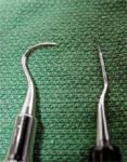
Figure 3. Hand scalers (left) have pointed tips, and curettes (right) have rounded toes.
Polishing tools
After hand or ultrasonic scaling, microetchings are present on the enamel. If left in this roughened state, the teeth may be prone to accelerated plaque and calculus accumulation. Polishing restores a smooth finish to the enamel. It is performed with a low-speed handpiece at a speed of 2,000 to 4,000 rpm, a rubber prophylaxis cup, and prophylaxis paste. Be sure to keep an ample amount of prophylaxis paste in the rubber cup because the paste polishes the tooth, and it also provides lubrication to reduce thermal damage. Mechanical toothbrushes are not a substitute for polishing.


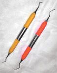
Figure 4. The Columbia 13/14 (left) and the Barnhart 1-2 (right) are two curettes commonly used in veterinary practice.
Dental radiography unit
Although the periodontal probe is an extremely sensitive instrument to assess periodontal disease, it does not provide the full clinical picture. Intraoral radiographs more accurately estimate the degree and character of perio dontal bone loss. Additionally, if tooth mobility is not due to periodontal disease, intraoral radiographs can shed light on the true problem. Intraoral radiography is superior to skull radiography for assessing the perio dontal tissues because it eliminates superimposition and provides better resolution. The dental radiography unit is relatively small and is installed in the dental operatory for maximum convenience (Figure 5). In addition to the radiography unit, dental film and appropriate developing equipment must be acquired. A digital dental radiography unit is an alternative to a film-based system.

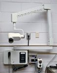
Figure 5. Standard wall-mounted dental radiography units with articulating arms are easy to position and give a detailed image of dental pathology.
EQUIPMENT FOR PERFORMING DENTAL EXTRACTION
In many cases, instruments specialized for veterinary patients will make the extraction process easier on both you and your patient.
Dental radiography unit
In addition to being a diagnostic aid, intraoral radiography is important for preoperative and intraoperative planning of dental extractions. Preoperative radiographs will help you determine how difficult the extraction will be and whether complications are probable. Intraoperative or postoperative radiographs are indicated to confirm the removal of all root fragments.
Periosteal elevator
Most dental extractions require developing a mucoperiosteal flap for ease of extraction and proper closure of the extraction site. A periosteal elevator is a flat, sharp instrument that is passed between the periosteum and bone to separate the mucoperiosteal flap from the bone. A variety of periosteal elevators are available, and the choice is based on the size of your patient and your personal preference. Two popular types, Molt and Freer, resemble flat spoons (Figure 6).


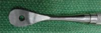
Figure 6. A Molt periosteal elevator for developing a mucogingival flap.
Wiggs winged dental elevators
Other styles of dental elevators are available, but we think Wiggs winged dental elevators are the most versatile for veterinary dentists. The working ends of these instruments are curved to better adapt to the conical shape of canine and feline teeth and come in a variety of sizes (Figure 7). The end should be kept sharp for cutting the periodontal ligament during extractions. Always remember to keep your index finger near the working end to reduce the potential of iatrogenic trauma to your patient such as orbital penetration.7


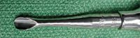
Figure 7. The working end of a Wiggs winged dental elevator. Note that the end is thin and slightly curved. This instrument should be kept sharpened for optimal use.
Small-breed extraction forceps
Many veterinary practices have an odd assortment of dental extraction forceps that are usually designed for people and are often inappropriate for veterinary use. They are often too large to use in veterinary patients and are also specialized for a particular tooth. Small-breed extraction forceps are versatile and small enough to use with most cats and small to medium-sized dogs (Figure 8). Forceps should only be used when the tooth has been properly elevated and is mobile. Forcibly extracting teeth with extraction forceps may lead to fractured roots or iatrogenic fracture.


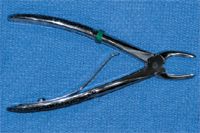
Figure 8. Small-breed extraction forceps are versatile and small enough for use in cats and small dogs.
High-speed handpiece or drill with burs
A high-speed handpiece or drill and a low-speed handpiece are both powered by air (Figure 9). The air source is either a compressor or nitrogen. Air pressure drives a small turbine in the high-speed handpiece, generating low torque but high speeds (200,000 to 400,000 rpm), thus driving the bur or bit. Burs for high-speed units are called friction grip burs and are designated by FG. When buying burs, do not confuse FG with RA (right angle) or HP (handpiece) burs because these will not fit the high-speed handpiece.

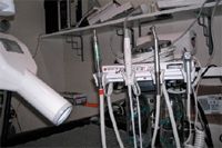
Figure 9. The components of an air-driven unit include (from left to right) a low-speed handpiece that can be used with a prophylaxis angle and cup for polishing teeth, an ultrasonic scaler (piezoelectric) that has been incorporated into this system, a high-speed handpiece for sectioning teeth and removing bone, and an air-water syringe for rinsing.
During extractions, burs are used to section teeth into individual root components and to remove the buccal cortical bone plate, making root elevation much easier. Burs are made of carbide and are either end-cutting or side-cutting. End-cutting burs, which are useful in removing bone, are either round (denoted by #1/4 through #8) or pear-shaped (#329 through #332). Side-cutting burs are useful for sectioning teeth and come in a number of styles. The most popular is the crosscut tapered fissure (denoted by #699 through #703). Although often used multiple times, carbide burs are designed for single use and are priced accordingly. Extended use leads to a dull, inefficient bur.
Water coolant should always be used to reduce thermal damage. It should also be noted that electric-driven handpieces are often marketed to veterinarians for the same use. These handpieces can be dangerous since too much torque is generated, which can lead to iatrogenic injury to the patient.
CONCLUSION
Some of the most commonly performed dental procedures in a veterinary practice are periodontal prophylaxis and dental extraction. When you have the right tools at hand, as described in this article, you can provide optimal dental care for all your patients.
Editors' note: Dr. Lemmons is a consultant for Veterinary Information Network. Dr. Carmichael has been a consultant for Pfizer, Henry Schein, Merial, and Webster.
The information and photographs for "Dental Corner" were provided by Matthew Lemmons, DVM, Circle City Veterinary Specialty and Emergency Hospital, 9650 Mayflower Park Drive, Carmel, IN 46032, and Daniel T. Carmichael, DVM, DAVDC, Veterinary Medical Center, 75 Sunrise Highway, West Islip, NY 11795.
REFERENCES
1. Wiggs RB, Lobprise HB. Periodontology. In: Veterinary dentistry principles and practice. Philadelphia, Pa: Lippincott-Raven, 1997;186-231.
2. Harvey CE. Management of periodontal disease: understanding the options. Vet Clin North Am Small Anim Pract 2005;35:819-836.
3. Holmstrom SE, Bellows J, Colmery B, et al. AAHA Dental Care Guidelines for Dogs and Cats. J Am Anim Hosp Assoc 2005;41:277-283.
4. Sanz M, Newman MG. Advanced diagnostic techniques. In: Newman MG, Takei HH, Carranza FA, eds. Carranza's clinical periodontology. 9th ed. Philadelphia, Pa: WB Saunders Co, 2002;487-502.
5. Wiggs RB, Lobprise HB. Dental equipment. In: Veterinary dentistry principles and practice. Philadelphia, Pa: Lippincott-Raven, 1997;1-28.
6. Brine EJ, Marretta SM, Pijanowski GJ, et al. Comparison of the effects of four different power scalers on enamel tooth surface in the dog. J Vet Dent 2000;17:17-21.
7. Smith MM, Smith EM, La Croix N, et al. Orbital penetration associated with tooth extraction. J Vet Dent 2003;20:8-17.