How veterinary medicine can save the world, Part 1: Curing disease
In the next few issues of dvm360, we're taking a close look at how veterinary medicine benefits people, not just animals. In this first installment, we meet a 'translational' (cross-species) researcher who's in the process of revolutionizing orthopedic medicine-for people and pets.
The pain never stops. It bites when you sit. It bites when you stand. It bites when you climb the stairs, push in the clutch, bend over to pick up a piece of paper. It even bites when you lie down and try to sleep at night.
When your orthopedist slaps the x-ray up on the wall and describes the situation as "bone-on-bone," you know exactly what the words mean.
They mean somewhere down the road, your knee joints or hip balls will be sawed off and replaced with plastic or titanium. Yes, titanium joints are beautifully machined to tolerances representing the best of what the human mind can engineer. And if all goes well and you do the rehabilitation work, you'll get relief from the pain—at least for as long as these devices last.
But what if there were a better way? What if those replacement parts could be living tissue? Better yet, what if the deterioration was caught early and your orthopedist started a regenerative process rendering implants unnecessary?
Those living tissue replacements are being tested today in the lab and the clinic, and if someday they come to a knee or a hip near you, you can thank a dog ... and a veterinarian.
Driven to help knees from the age of 5
James (Jimi) L. Cook, DVM, PhD, DACVS, DACVSMR, holds dual appointments in veterinary and human medicine at the University of Missouri in Columbia, Mo. On this particular morning he's in the process of moving his office. His dual appointment has been 75 percent in the veterinary school and 25 percent in the medical school, but now the proportions have reversed and his new office will be a few long strides away in the medical building.

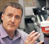
Dr. James Cook of the University of Missouri holds a vial containing orthopedic donor tissue stored in a special fluid. The preservative, which he helped develop, keeps donor tissue healthy for two months, compared to the more traditional 30-day window. This means less wasted tissue.
Cook heads a team of 30 colleagues who work in two labs, one biologic and one engineering, both under the umbrella of the Comparative Orthopaedic Laboratory (COL) at Missouri. Its motto: "Finding joint solutions." Cook has been searching for joint solutions in dogs since he entered Mizzou's PhD program in the mid-1990s, always with the aim of helping their human counterparts.
In fact, Jimi Cook has been driven since the age of 5 to do something about the pain in human knees. In 1975, his grandfather, Robert B. Gordon, had just undergone knee surgery and the prescription for rehabilitation was bike riding. So every morning he took his grandson out for a spin.
"I became part of his rehab," Cook says. "We'd ride bicycles every morning. We'd talk about everything under the sun on those bike rides. In my mind we were trying to solve this arthritis problem with my grandfather."
The memory is bittersweet because his grandfather's arthritis was a constant battle between science and pain. A tennis player and water skier, Gordon had severe primary degenerative arthritis in both knees. His first surgeon implanted a pair of pound-on prostheses, which Cook still has in his office. It's not hard to imagine how a researcher holding those metal wedges in his hands might be driven to produce true replacements from living tissue.
"When I'm speaking around the world I ask the people in the crowd to hold up their hands if they would love to have metal or plastic parts in their joints," Cook says. "I say, 'Raise 'em up real high and keep 'em up.' And, of course, no one ever raises their hand, anywhere I've been. So, while metal and plastic are better than the alternative, they're not perfect. There's pain, rehabilitation, complications. Even if the surgery goes well, honestly, you're still limited in function. No orthopedic surgeon will tell you that after your knee is replaced you're going back to what you were doing before."
Seven additional surgeries were doubtless not what his grandfather envisioned either in the mid-1970s when he trusted his knees to his first surgeon. Gordon was, in Cook's words, a self-made man who rose from sweeping floors at Airetool Manufacturing to president of the company. He wasn't the sort to give in to disease; instead he started researching the journals. He found an article in Scientific American describing the pound-on procedure. Cook has a rendering of the magazine cover and his grandfather's hand-written notes in the margins on a plaque. "I think he may have been the fifth person in the country to have that surgery," Cook says. "It was still very rare."
Pioneering as the procedure was, and as helpful as the seven additional surgeries were—certainly preferable to a wheelchair, amputation or joint fusion—Gordon still had skin grafts and infection, leg length and loosening—"all the problems associated with metal and plastic," Cook says.
Much like his grandfather, people hear about what Cook is doing at Missouri and write him letters desperate for the latest advances. He puts them on a list that now numbers over a hundred. "They say, 'I'll sign any release you want me to sign; I'll fly you to Europe or China or wherever you can do it.' And I say, 'I'm a veterinarian,' and they say, 'I don't care. I don't want metal or plastic. I want you to do it. I'll sign any release. I'll pay any amount of money.'"
The veterinary side of the equation
People aren't writing Cook just for the pain in their own knees and hips. They're bringing their dogs to the clinic—sometimes in a last-ditch effort—for live tissue implants and other advanced procedures in hopes of getting them back to work or back in the field.
Cook's team works with all kinds of working dogs: agility, search and rescue, military, police, hunting, service and field trial dogs. Last year Cook treated an elbow in a rescue dog using a Canine Unicompartmental Elbow (CUE) developed in the Missouri lab for Arthrex VetSystems. The dog, Lincoln, hadn't worked for more than a year when Cook implanted the living cartilage using the CUE medial resurfacing procedure. Lincoln passed his six-month checkup in early May and Cook got word he went to Moore, Okla., to help with the rescue operation after the massive tornado of May 20th. "He helped find some people and some bodies," Cook says. "That's pretty cool."
One particular field trial dog has become a staple of Cook's talks, a symbol of what can be done with live tissue and a key link to human applications. His name is Buddy and he was 7 years old at the time of the surgery. Cook opens the silver Mac on his desk, clears away clutter from his office move, and quickly finds the video record of Buddy's recovery. The first video shows Buddy unable to put his left leg down even with the buoyancy of water on the underwater treadmill. He looks nothing like the athlete who had won field trials and become a favorite of his owner and trainer. Buddy had been subjected to four knee surgeries, the last of which removed both menisci. His whole countenance, Cook says, his appearance and his mood, had degenerated. His owner read about the work Cook was doing at Missouri and decided to give Buddy one last chance.
"He was completely non-weight-bearing lame," Cook recalls. "His owner said, 'Listen, doc, I love this dog but this dog is miserable.'" Buddy had lost 15 pounds and, in Cook's estimation, was depressed. "Those dogs live to work," he says. "If they can't work, literally, they will not eat."
The owner told Cook, "I'm going to have to put him to sleep. Not because I don't care about him, but because I care about him too much. He can't live like this."
Cook explained that the procedure had been effective in research dogs and was being considered in human medicine but was not approved yet. The owner decided to give it a try. So Cook completely replaced Buddy's knee joint with cartilage, bone and menisci grown in the Missouri lab from organ donor grafts. In his clinic office today, three years later, Cook pushes the play button on the Mac and the video shows Buddy "running like the wind," in Cook's words, across an open field, cutting and maneuvering like a halfback slicing through the line. Buddy's even back to winning field trials, Cook says, and he's passed all his checkups.
"I've shown this video all over the world," Cook says. "Every time I watch it—and I've shown it literally hundreds of times—I get goosebumps."
Buddy wasn't the first dog to have live tissue implants at Missouri. The first was a dog from Minnesota who received an implant seven years ago, and the elbow is still sturdy. Dr. Cook recently had two dogs in the clinic with biologic joints, and a third, from Vancouver, British Columbia, had been discharged the day before after a cartilage graft.
The cartilage graft is interesting because that procedure originated on the human side, Cook says. "So," he says, "it comes full circle."
Building bridges for dog and man
Medical research has long been stranded on a set of islands. On one island lived the bench researcher in pursuit of the secrets of pure science. On another lived the clinical researcher who waited years, even decades, for pure research to reach the application phase. And, of course, the veterinary researchers lived on their own islands, apart from their human medicine counterparts.
Since money is almost as critical to research as ideas, all of those islands were fighting for pieces of the same pie.

10 ways researchers are studying diseases in dogs and humans
Today, that's changing. Bridges between islands are under construction in labs like Cook's at Missouri. Bench research is reaching clinical researchers more quickly. And veterinary medicine is reaching out to human medicine even as human medicine reaches out to veterinary medicine. "I know this is a cliché and I apologize," Cook says of the translational research paradigm, "but this is simply a win-win proposition for everybody."
For example, veterinary researchers have long been cramped by an economic ceiling on their efforts. To win funding for a project, they must demonstrate the potential for human application.
"It's just a dead, straight-on fact that funding is going to go toward human medicine," Cook explains. "What's cool is that we can leverage that funding to help canine patients. It's there for the humans, we test it in the dogs, but we can also apply it safely and effectively to the dogs. We're healing the species that we've developed this product through."
Cook adds that some major human medical firms are now investing in veterinary applications as a result of the research. In fact, he says, Arthrex now has a veterinary division based on some of the research originating in his lab and honed in the clinic. "They've seen what it can do in the marketplace in the real world on the veterinary side," Cook says.
At Missouri, the veterinary side and the medical side have grown closer across the past decade. Cook says one reason is simple proximity; it's just an eight-minute walk across campus from the veterinary building to the medical facility. His team includes engineers, veterinarians, molecular biologists and medical doctors. Interaction between the veterinary side and the medical side happens weekly if not daily. The new department chair in orthopedic surgery, James P. Stannard, MD, he says, clearly understands how research on the veterinary side can make the university "the best in the world" on the human side.
The bridge between veterinary medicine and human medicine being built at Missouri began with a plan Cook sketched out with Keith Kenter, MD, in 1995 on a napkin at Buckingham's Smokehouse Bar-B-Q in Columbia. Kenter was a medical doctor at roughly the same stage in his career as Cook. Kenter has since moved to the University of Cincinnati, but their napkin-based plan to combine veterinary research with human applications has grown from a lab "the size of a closet" in 1998 to a multimillion-dollar operation. "It grew quicker and bigger than I ever imagined," Cook says. "We found passionate people who are team players and—we always say—teams defeat individuals."
Cook says a typical research question starts with a question: "What can't you tell a patient today?" "The answer might be that I can't replace their joints with biological tissues," Cook says. "I can't tell a patient he or she can go back to full function. Once we know the question, we put the science behind it."
Then the research goes from the laboratory through an animal model for safety and testing. Most of the time, for Cook's lab, the animal model is a dog. Then, via a long pathway with the FDA, researchers progress to human clinical testing and use. But in the meantime, canine patients directly benefit through clinical application.
The big question: Why did it take so long to join forces?
Today, the synergies between veterinary medicine and human medicine seem obvious. Several products of Cook's lab are already approved for humans. When the FDA approves living tissue joint replacements, which Cook says will likely take about seven years, the connections will be even more evident to the lay public. And, he says, the evidence most persuasive so far to the FDA has been his videos of Buddy running in the field. So why has it taken this long to build bridges between the veterinary and human research islands?
"The first-blush answer is that we didn't realize that it was one medicine, one health," Cook says. "When you just look at a cow and a human you automatically see there are differences—four stomachs in a cow comes immediately to mind. I think our tendency has been to look at the differences first instead of the similarities. And to stop there."
But canine knees, hips and elbows, it turns out, are excellent models for human joints. Cook finds another set of images on the silver Mac then turns the screen around. The first image is a pair of still images of two open knee joints. The second is a pair of videos of orthoscopic meniscus repair. "I tell people, one of these is a dog and one of these is a human. If I was mean, I'd make the audience tell me which one is which. This is the human knee," he says, pointing to the image of the right. "This is the dog knee. You can see they are almost identical." The arthroscopic videos, he says, are even more difficult to distinguish, so much so that even some orthopedists can't tell the difference.
So the problems in dogs are the same and the treatments are the same, Dr. Cook says. Even the rehabilitation procedures have turned out to be the same. These similarities are more obvious, he says, where working dogs and canine athletes are concerned. With 2.5 million registered agility dogs, mobility problems have become more obvious, paralleling mobility problems in human athletes. Owners notice subtleties: The dog ticks the bar when he used to clear 20-inch jumps easily or refuses the A-frame twice before he agrees.
"It's funny that it has taken so long," Cook says, "because we've anthropomorphized everything else about dogs, everything from diet to emotions to car seats. Everything but the medical parts."
He says the wide distribution of information made available by the Internet has also hastened bridge building, each side becoming more aware of what the other is investigating. And financial pressures also play a key role as medical firms and granting institutions seek ways to maximize research dollars by pushing harder for clinical applications of pure science.
"I think what's cool about it is the reaching out, the bridging, is coming from both sides today," he says. "It's not veterinarians going over and begging at the doors of the MDs. Or vice versa. Not MDs coming in saying, 'Please work with us, please do animal models.' It's really the realization that what we are doing is very similar."
Have cultural differences between veterinarians and medical doctors also stood in the way?
"I think 'cultural' is probably the most polite way to put it," Cook says. "I think there was the veterinarian feeling inferior at times and the MDs maybe perpetuating that. But I think that's starting to bridge, too. If you had to go span the gap by yourself, it would be intimidating. I've been blessed at Missouri. People have respected me sometimes more than I deserve."
Walking down the hall to his biologic lab with the long, sure strides of a man who was once a professional water skier, Cook describes the similarities between dog and man and the realization of what a good research model the dog is for human orthopedic medicine. As he turns the corner, he finishes the thought.
"When I'm under the drape, I don't care whether it's a four-legger or a two-legger," he says. "I want to fix what's wrong. And, I want to fix it better."
John Lofflin is a freelance writer in Kansas City, Mo., with extensive experience writing about the veterinary profession.
Podcast CE: A Surgeon’s Perspective on Current Trends for the Management of Osteoarthritis, Part 1
May 17th 2024David L. Dycus, DVM, MS, CCRP, DACVS joins Adam Christman, DVM, MBA, to discuss a proactive approach to the diagnosis of osteoarthritis and the best tools for general practice.
Listen