Part 1: Veterinary dental radiography: a vital practice resource
Two case examples display how radiography can reveal hidden pathology.
High-quality veterinary dentistry is impossible without the diagnostic tool of dental radiography. The extreme importance to patient care of full-mouth radiography will be demonstrated in this series of articles.
The results of two prospective nested case-control studies conducted at the University of California-Davis are worth a close look. Researchers found that of 226 dogs studied, dental radiography of teeth without clinical lesions demonstrated clinically significant lesions in 27.8 percent of dogs.1 In a similar study involving 115 cats, radiographs of teeth without clinical lesions nonetheless demonstrated clinically significant findings in 41.7 percent of subjects.2
What diagnostic modality currently present in your practice has this percentage of significant positive results that require treatment? See the importance of dental radiography yourself with these two case examples.


Case 1
A 5-year-old spayed female miniature schnauzer was presented for routine oral evaluation and professional teeth cleaning. The only abnormality noted was a missing first premolar on both mandibles (Photo 1). Radiography revealed bilateral dentigerous cysts associated with the unerupted teeth (Photo 2).
Dentigerous cysts are classified as odontogenic cysts and arise from the epithelial components of the developing tooth follicle or remnants thereof.3 Normally on eruption, the follicle becomes the junctional epithelium, which is present at the base of a normal tooth sulcus. If the tooth doesn't erupt, this epithelium has a strong tendency to produce cysts.
Dentigerous cysts are a common finding when teeth are missing, especially the mandibular first premolars. Brachycephalic and small-breed dogs seem to be predisposed. The mandibular first premolar is not the only tooth whose absence should prompt suspicion. Any areas of either jaw that are missing teeth should be evaluated radiographically in all breeds of dogs and cats to determine whether cysts are present. In my experience, most teeth that are unerupted will form cysts. Treatment involves referral to a veterinary dental specialist (avdc.org) for surgical exposure and complete removal of the tooth and cyst (Photos 3 and 4).
Case 2
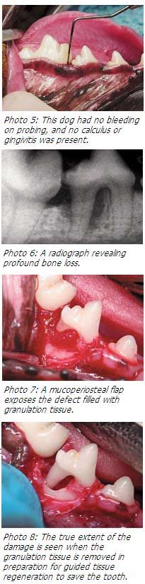
A 4-year-old neutered male dachshund was presented for routine oral evaluation and professional teeth cleaning. No bleeding was present on probing (Photo 5). However, the radiographs showed tremendous bone loss adjacent to the distal root of the left mandibular fourth premolar (407), evidenced by the dark area around the roots (Photo 6). The lucency does not represent a void but rather a mass of infected granulation tissue (Photo 7). The true extent of the damage was evident once the diseased tissue was removed (Photo 8).


Although this tooth could be saved by veterinary dental specialists, daily brushing and alternative home care such as special diets, chews and water additives (products approved by the Veterinary Oral Health Council can be found at vohc.org) are prerequisites when making the decision not to extract. Frequent professional cleaning every three to six months is a required pet guardian commitment.
Conclusion
These case studies are just two examples of the power of veterinary dental radiography. Keep in mind that the studies mentioned earlier looked at clinically normal teeth. Imagine the hidden pathology of all of your current patients that have tartar and gingivitis.
More on the tremendous impact that dental radiography can have for your patients and your practice will be examined in the months to come.
Dentistry by Brett Beckman DVM, Dipl. ACVD, Dipl. AAPM
Dr. Beckman is acting president of the American Veterinary Dental Society and owns and operates a companion-animal and referral dentistry and oral surgery practice in Punta Gorda, Fla. He sees referrals at Affiliated Veterinary Specialists in Orlando, Fla., and at Georgia Veterinary Specialists in Atlanta, lectures internationally and operates the Veterinary Dental Education Center in Punta Gorda, Fla.
References
1. Verstraete FJ, Kass PH, Terpak CH. Diagnostic value of full-mouth radiography in dogs. Am J Vet Res 1998;59(6):686-691.
2. Verstraete FJ, Kass PH, Terpak CH. Diagnostic value of full-mouth radiography in cats. Am J Vet Res 1998;59(6):692-695.
3. Wiggs RB, Lobprise HB. Clinical oral pathology. In: Veterinary dentistry: principles and practice. Philadelphia, Pa: Lippincott-Raven, 1997;131.