How to know if your veterinary hospital needs a CT scanner
Thinking of buying this advanced imaging technology for your general-practice veterinary hospital? A veterinary imaging specialist answers the money, time and space questions.
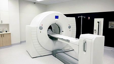
Yes, they make models of veterinary CT scanners smaller than this. (WADII / stock.adobe.com)

Computed tomography (CT) has proven beneficial in all aspects of veterinary medicine, including surgery, oncology, dentistry and internal medicine. Not only can it provide an imaging diagnosis (such as the presence of pulmonary or nasal masses), but you can obtain CT-guided aspirates and biopsies for a cytological or histopathologic diagnosis. Owners are asking more frequently about CT for their pets, and more veterinary specialty and general practice hospitals want to be able to offer this service to their clients.
General practitioners have a very challenging specialty of their own in that they deal with all aspects of veterinary medicine and are often the front line of care for patients. So … do general practitioners need to offer in-house CT scans?
What's it worth to you?
A CT scanner can be a big investment in money, training, maintenance and space. For fixed CT scanners, the room-including the glass-must be shielded, which adds to the initial cost. Will the hospital be doing enough CT scans that it will be profitable for their business?
What do you know about it?
Another consideration is the veterinarian's knowledge of CT: proper protocol and positioning, along with the correct indications for and limitations of the technology. The clinician should also be able to identify artifacts and troubleshoot the CT during the scan if needed.
No matter who's interpreting, the quality of the study will greatly influence its diagnostic value. Unlike in human medicine, most veterinary patients are anesthetized for their CT scan, so you need to try to get it right the first time. For example, most thorax scans should be done with a breath hold to decrease atelectasis. If there is pleural effusion, the pet may need a thoracocentesis before the scan if it's safe to perform one.
Deeper details: Cone vs. slice CT
In cone-beam CT, a cone-shaped X-ray beam is detected by a flat-panel sensor. Both the sensor and X-ray beam rotate around the patient, creating a larger volume of data in a rotation.
In slice CT, a narrow beam rotates around the patient and is detected by an array panel sensor, creating a series of thin slices.
Because a cone beam creates a single larger volume of data that's then reconstructed, it's more susceptible to motion artifact than a slice CT. If there's transient motion, it will affect all of the images. A cone-beam CT also rotates more slowly around the patient, making it inherently more susceptible to motion artifacts.
With slice CT, only the slices that have motion will have artifacts.
Slice CT emits collimated X-rays in thin slices through the patient that are detected by the opposing detector.
Because cone-beam CT scans a larger section at once, there's less collimation and more stray X-ray beams get registered on the plate. As with radiographs, the bigger the body part imaged, the more scatter. So, scatter would be higher for a large dog abdomen or thorax compared to a cat or a limb. The increase in scatter leads to decreased contrast resolution (the ability to distinguish different shades of gray).
Advantages of cone-beam CT include decreased radiation dose to the patient, decreased cost to purchase the unit and better spatial resolution (the ability to distinguish two points from one another) compared to slice CT. Also, cone-beam CT takes up less space than a fixed-slice CT scanner.
How big is a CT scanner?
In general, a fixed multislice CT scanner needs to be housed in a room that's at least 14 by 20 feet, according to my source at Sound. However, this can vary depending on the size of the machine. A control area outside of the room is also needed for the technician to be able to run the scan. The room-including the glass window-must be shielded with lead that's formulated by a physicist or someone trained in radiation shielding. This depends on the specific CT scanner and its proximity to other rooms and personnel as well as other considerations.
There are also smaller portable multislice CT systems that require less space, and some of these require no shielding at all. These portable CT scanners were developed for use in human medicine for emergencies when a patient might not survive transport to a facility with a fixed CT. Some of these CTs, however, have a smaller gantry size that limits the size of the patient that can be imaged. Talk to your vendor for more information.
Would you rather refer?
Although not all specialty hospitals have a radiologist, there are benefits to having CT done in a facility with an on-site radiologist and other specialists. A radiologist can evaluate the images during the scan while the patient is still under anesthesia, and aspirates or biopsies of abnormal lymph nodes or other tissue can be obtained under the same anesthetic event.
Patients often have procedures immediately after the scan-if the patient has a nasal mass, for example, an internist could go straight to rhinoscopy with biopsies. Depending on the comfort of the clinician, a veterinarian could see a reduction in the number of anesthetic events as well as the time it takes to reach a diagnosis if a patient is referred for CT scan.
Before you dive in and buy a scanner, I'd encourage you to reach out to a veterinary radiologist or spend some time at an imaging facility with a radiologist to become familiar with the protocol, positioning and indications for CT. This will help you make the best decision for your practice-and provide the best diagnostic scan for your patients.
Dr. Ketaki Karnik is an associate at Southern California Veterinary Imaging in Culver City, California.

Celebrating veterinary technicians
Credentialed professionals share what makes them feel appreciated in their role and talk about National Veterinary Technician Week
Read More