The ABCs of veterinary dentistry: "I" is for informed consent
An in-depth look at what can go wrong during a dental procedure and how much to share with veterinary clients.
My sister asked me to accompany her to a surgical consultation for an elective procedure. After the doctor finished reviewing the proposed surgery plan he said, “Now let's talk about what can go wrong,” listing infection, dehiscence, suture reaction, scar formation and significant postoperative discomfort, which rarely occurs. He followed with, “Now what questions or concerns do you have?”
Side consideration: Maybe you should refer?
Mollie, our 15-year-old poodle.
Twelve years ago, our 3-year-old standard poodle began vomiting, accompanied with bloody diarrhea. These signs continued despite symptomatic care. Radiographs revealed intestinal obstruction. My brother-in-law, a human colorectal surgeon, volunteered to scrub in with me. Together we performed the exploratory to reveal a volvulus with greater than half of her small intestines purple.
The choices were to resect a major amount of her small intestines right then and there or to close her up and refer to a surgical facility 10 miles away. I chose to refer. She was reopened, the surgeon placed a tube for decompression into the area and did not have to resect any intestine. Our dog stayed at the referral hospital for a week, and 12 years later is still very much alive. In fact, she just celebrated her 15th birthday.
What does this have to do with dentistry? Consider referring your tough cases. When you do not feel comfortable proceeding either before the case begins or during if a complication occurs, refer. If you are willing and able, go with your patient to the veterinary dentist. Everyone comes out ahead.
Why did he do that? To worry us? To talk her out of the procedure? Not at all. He wanted to properly inform us-and in some way to be brutally honest that things do not go perfectly every time. The informed discussion and my sister's consent was founded on her right to make health decisions based on explanation and understanding the risks and benefits of treatment and nontreatment. Immediately after the visit, I consciously incorporated “Now let's talk about what can go wrong …” during all client discussions on proposed surgeries or diagnostics that carry risk.
For a general physical examination, radiography, electrocardiography (ECG) and blood draws, implied consent is assumed. For diagnostics and treatment with risk or alternatives, we need to give our pet owners enough information for them to make informed diagnostic and therapeutic decisions. Blindly signing a consent form without reading and discussing content is not ethically adequate. Informed consent needs to be a communicative process, presented several times during the professional oral assessment, treatment and prevention (oral ATP) visit.
This informed discussion should include:
1. The nature of the procedure
2. Reasonable alternatives if any
3. The relevant risks, benefits and uncertainties related to each alternative
4. Confirmation that the client understands the above
5. The acceptance of the decision and procedure by the client
Deciding how much “what can go wrong” information to give the pet owner can be challenging. Fortunately, adverse events rarely occur. For example, a dog or cat hardly ever loses its hearing-even temporarily-from acoustic nerve trauma caused by the ultrasonic scaler. Even though it does happen, it has never clinically occurred in one of my patients. But it could, so do I discuss it beforehand? No, because it is rare and not life-threatening. Compare this to adverse events that can occur secondary to general anesthesia. Even though these are also extremely rare, they can be devastating. For that reason, we discuss anesthesia informed consent with every case.
Examples of common informed consent topics to consider discussing relating to dental procedures
General anesthesia
-Adverse anesthesia events including death
Probability: Extremely rare
Prevention:
1. Evaluate the patient beforehand with a physical examination, laboratory testing, ECG, ultrasonography and radiography, as indicated.
2. Choose an anesthetic protocol tailored to the patient.
3. Monitor vital signs during and after the procedure.
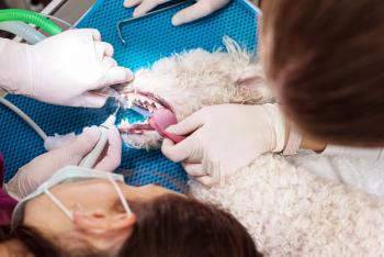
4. For compromised patients or intense clients, consider using a veterinary anesthesiologist or referring to a facility with one (Figure 1).
Figure 1. A veterinarian dedicated to tailor anesthesia protocols, monitor effects and make anesthesia modifications during the procedure. (All photos courtesy of Dr. Jan Bellows)
-Coughing
Probability: Moderate to high
Prevention:
1. Use sterilized, appropriately sized endotracheal tubes inflated to proper cuff pressure.
2. Avoid excessive head movement during the procedure.
3. Disconnect the endotracheal tube attachment from the anesthesia machine when the patient is moved from side to side.
Tooth extractions
-Excessive bleeding
Probability: Moderate with extractions
Prevention:
1. Appreciate vascular anatomy before any extraction.
2. Raise the patient's head and apply hemostatic powder to control excessive bleeding (Figure 2).
3. Consider referral in cases of persistent excessive bleeding at an extraction site with root fragments remaining.
Figure 2. Hemostatic powder applied to stop hemorrhage from a fourth premolar extraction site.
-Postoperative discomfort
Probability: Moderate
Prevention: Provide local and systemic anesthesia and analgesia before, during and after a procedure in which pain may be expected.
-Jaw fracture
Probability: Extremely rare
Prevention:
1. Obtain radiographs of all teeth and surrounding tissues before extractions.
2. Use your surgical expertise as well as patience, illumination and magnification, especially in cases of advanced periodontal disease extending to the mandibular ventral cortex.
3. Consider referral if you are not comfortable with the surgical extraction-or refuse to do the procedure if you feel that jaw fracture is a significant risk (Figure 3).
Figure 3. A radiograph of a difficult extraction of the left mandibular first molar, potentially leading to jaw fracture.
-Surgical site infection, dehiscence
Probability: Rare
Prevention:
1. Explain to the client that sometimes after surgery a dog or cat opens its mouth too far and sutures fail to hold tissues together. Usually the surgical site then heals by secondary intention.
2. Create a flap that will allow closure without tension.
3. Consider sending a patient home with a cone to decrease oral cavity self-trauma.
-Remaining root fragments after surgical extraction
Probability: Rare to moderate
Prevention:
1. Obtain and examine intraoral radiographs on all teeth to be extracted before and after the procedure.
2. Refer tough cases if you are not comfortable, such as multirooted teeth and tooth resorption (Figure 4).
Figure 4. A root fragment remaining from an extraction of the maxillary fourth premolar.
-Oronasal or oral antral fistula
Probability: Rare to moderate
Prevention: Prepare a gingival flap to facilitate closure without tension. Let the client know that if a fistula occurs and becomes clinically significant, surgical closure is advised (Figures 5A and 5B).
Figure 5A. An oral nasal fistula.
Figure 5B. An oral antral fistula.
-Anorexia
Probability: Moderate for one to two days
Prevention:
1. Provide pain relief as well as anti-inflammatory and appetite-stimulant medication.
2. In cases of full-mouth extractions in debilitated cats and other major oral surgical procedures, place a nasogastric or pharyngeal feeding tube (Figures 6A and 6B).
Figure 6A. A nasogastric feeding tube.
Figure 6B. Pharyngostomy feeding tube placement.
-Tongue protrusion after multiple incisor or quadrant extractions
Probability: Moderate
Prevention: Let the client know that tongue protrusion has little to no effect other than cosmetic (Figures 7A and 7B).
Figure 7A. Tongue protrusion after surgery was performed to remove the rostral mandibles from the first molars forward in the patient pictured in Figure 3.
Figure 7B. A right-sided tongue protrusion after rostral mandibulectomy to remove an invasive oral melanoma.
Oral mass removal
-Dehiscence
Probability: Moderate
Prevention: Close without suture line tension.
-Regrowth
Probability: Common with incomplete excision
Prevention:
1. Perform fine-needle aspiration cytology before surgical excision to help determine malignancy and plan surgical margins.
2. Perform computed tomography or cone beam computed tomography before surgery to plan surgery with clean margins (Figures 8A-8D).
Figure 8A. An oral mass involving the rostral maxilla.
Figure 8B. Removal of the mass (osteosarcoma) with clean distal surgical margins only.
Figure 8C. Three-month rostral regrowth of the tumor.
Figure 8D. Five-month continued regrowth.
-Possibility of further oncologic treatment needed
Probability: Moderate in cases of malignancy
Prevention: Before surgery, mention to the client the probability of radiation therapy in those masses that are responsive.
Consider discussing these very rare adverse outcomes with clients who you think need to know more:
General anesthesia
-Vision loss
Probability: Extremely rare
Usually attributed to anoxia or very low blood pressure during or immediately after the procedure.
-Lameness
Probability: Rare
Older animals and those with disk disease or arthritis may become lame secondary to prolonged positional changes.
-Trachea rupture, subcutaneous emphysema
Probability: Rare, primarily in older cats
To prevent this complication, be careful not to overinflate the endotracheal cuff, and disconnect the endotracheal tube every time the animal is rotated (Figure 9).
Figure 9. Pneumomediastinum, pneumothorax, and pneumoperitoneum secondary to tracheal rupture.
Ultrasonic scaling
-Hearing loss
Probability: Extremely rare
To prevent this complication, avoid the “jack hammer” effect by using the side of the ultrasonic scaler vs. the tip to remove plaque and calculus, and only spend seconds on each tooth; if more time is needed come back after addressing other teeth.
-Tooth discoloration secondary to pulpitis
Probability: Rare
To prevent this complication, avoid excessive heat transfer from the ultrasonic scaler by ensuring proper water spray, and only spend seconds on each tooth; if more time is needed come back after addressing other teeth.
Extractions
-Air embolism from the dental drill
Probability: Extremely rare
To prevent this complication, try to avoid entering the mandibular canal during the extraction process. An air embolus can be created by inadvertent injection of a mixture of air and water through the dental drill directly into the mandible entering the superior vena cava and right atrium.
So, how much information to share?
We should include a similar discussion as my sister's surgeon before dental procedures requiring general anesthesia. Veterinarians are held to reasonable community standards. What would a typical doctor disclose? Tell them what a typical client needs to know. It's just good practice and part of our practice acts. Of course there is a fine line between informing and scaring clients out of needed care. This is where the art of practice comes into play. Let your client know that these complications are for the most part extremely rare and you and your staff take every precaution to prevent them. Also convey the small risk is worth the reward of a pain-free functional mouth.
Newsletter
From exam room tips to practice management insights, get trusted veterinary news delivered straight to your inbox—subscribe to dvm360.


