Bronchopulmonary disease in cats-is it really asthma? (Proceedings)
Feline bronchial disease goes under a multitude of names reflecting the considerable heterogeneity in anatomic locations as well as etiologies that may be involved.
Feline bronchial disease goes under a multitude of names reflecting the considerable heterogeneity in anatomic locations as well as etiologies that may be involved.
Persistent tracheobroncheal irritation causes the chronic (>two months) cough that is easily elicitable on tracheal palpation, the hallmark of chronic bronchitis. Morphologic changes occur in the tracheobronchial epithelium and wall, resulting in airway inflammation: mucosal edema and thickening, epithelial metaplasia and cellular infiltrates with an increased production of mucus. This mucus is extremely irritating, resulting in a self-perpetuating process. When the irritation is occurring mainly in the small airways that are less than 2 mm in diameter, rather than cough being the main clinical finding, an expiratory wheeze predominates. As the airways become obstructed, the air becomes trapped within the alveoli resulting in a chronic obstructive pulmonary disease (COPD), with an abdominal thrust component to the respiratory pattern and, over time, resulting in a caudal displacement (or flattening) of the diaphragm. This patient may present in dyspnea with cyanosis rather than being brought in with a history of nocturnal wheezing or coughing. Clients may bring their cat in because of post-tussive retching or gagging misinterpreted as vomiting. It is the irreversibility of early changes at any stage along with the inherent progression that makes conclusive diagnosis and ongoing treatment so important.
From a different perspective, coughing may be due to tracheobronchial irritation, mucus secretion, accumulation, and bronchoconstriction. The result of this is airway narrowing, increased airway resistance, decreased air-flow, air exchange, hypoxemia, exercise intolerance, dyspnea with an increased respiratory rate. Potentially cor pulmonale or pulmonary hypertension could develop. The use of anti-tussives, is, however, contraindicated as the coughing itself is not detrimental and antitussive agents thicken secretions making it more difficult to clear the mucus.
Etiology
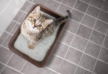
Despite the fact that we may call bronchopulmonary disorders "allergic bronchitis", the underlying etiologies and mechanisms are unknown. There has not yet been evidence to show that the clinical signs are associated with an increase in pulmonary mast cells, histamine release or IgE production. The presence or absence of eosinophils in secretions is not adequate proof that the condition is "allergic" in origin. Numerous inflammatory conditions/agents may incite the condition. These may include viral respiratory tract infections or inhaled irritants such as dust, aerosols, second-hand smoke, incense, hoods on litter boxes trapping dust, forced air heating/cooling, open fireplace use, use of sprays (pesticides, carpet cleaners, perfumes, deodorizers, oil misters). Alternately, these may be "triggers" acting to cause exacerbations of a chronic underlying condition.
Signalment
Classically it is the young adult cat who is affected with a preponderance of the Siamese breed being represented; however, any age or breed may develop bronchopulmonary disease.
History
Signs range from chronic coughing, retching or expiratory wheezing, (especially at night), to severe respiratory distress and cyanosis. It is important to note that cats can adapt to their disease state. Thus, any further compromise may be critical. This explains why some clients are not aware that their cat has been ill when they present for a "first" acute attack. Vomiting may be misinterpreted by clients when they witness retching after severe coughing, or may be secondary to aerophagia (from respiratory distress or pain) or gastrointestinal disease.
Physical examination
In most instances, the patient will be afebrile, bright and alert. Normal breath sounds may be present in mild cases; an expiratory wheeze may be ausculted over the lungs as the cat is trying to force air out of the smaller airways. This may be so marked that there is obvious abdominal thrust visible. Palpation of the trachea often results in a cough. As this loosens the mucoid secretions in the airways, post-tussive auscultation may detect some crackles. Note: if no breath sounds are heard at all in a patient, this reflects severe bronchoconstriction; this cat is probably cyanotic.
Differentials
As a general "rule-of thumb", changes in inspiration reflect an upper, larger airway problem, whereas, those of expiration reflect smaller, lower airway disease. Because of increased tracheal sensitivity in most cats, one may also need to consider upper airway obstructions due to laryngeal paralysis or masses, nasopharyngeal polyps, severe rhinitis or nasal obstruction. Anything that causes inflammation due to pressure or the presence of fluid as a result of some other disease process should be considered.
Diagnosis
If a patient is cyanotic or in severe respiratory distress, oxygen supplementation and rest are indicated before attempting to take radiographs or pursue other diagnostics. A small dose of a narcotic, such as oxymorphone, may help the cat to relax and thereby aerate his/her lungs more effectively. An oxygen cage, nasal oxygen or oxygen hood may be used. It is important not to use diuretics unless sure that the patient has cardiogenic pulmonary edema, as diuretics inspisate secretions and make them harder to clear from the airways.
Radiography
Once the patient is stable enough, thoracic radiographs are the indicated. Ideally, three views should be taken: right and left lateral and VD. The upper lung fields are less compressed and are, as such, viewed more clearly than the dependent lobes. Characteristic findings include a bronchial pattern; consolidation of right middle or caudal left lung lobes may occur from bronchial obstruction with mucus and debris with subsequent resorption of residual alveolar air; varying degrees of air trapping. If unresolved, eventually air trapping results in chronic over inflation of the lungs with diaphragmatic flattening. Sometimes alveolar or interstitial infiltrates may be seen. Occasionally, cats with broncho-pulmonary disease show no radiographic changes. Radiographic assessment of the heart should be made and, if there is indication of cardiovascular disease, followed up with an echocardiogram or, if there is an arrythmia, an ECG.
Bloodwork
Hematological and biochemical changes may be non-specific. Stress or bacterial infection may cause a neutrophilia. A peripheral eosinophilia may support the diagnosis of bronchitis, however may be from another cause (parasitic, hypersensitivity): the lack of such a finding does not rule out airway inflammation. Hyperproteinemia may be present. Heartworm antigen tests are indicated in endemic regions.
Fecal examinations
The eggs of Paragonimus or Capillaria may be seen in fecal examination: the former agent may be isolated using a sedimentation technique, while the latter may be seen with routine fecal examination. Aelurostrongylus larvae are detectable using a Baermann flotation technique.
Airway sampling
Two methods are commonly used to harvest airway secretions for cytologic and microbiologic evaluation analysis for differentiation and diagnosis of the various causes of coughing and/or wheezing in the cat. Tracheal wash is readily available to all practitioners and samples the contents of the larger airways. Using a sterile endotracheal tube is less stressful than the traditional trans-tracheal technique. Prior to insertion of the endotracheal tube, desensitize the glottis well using lidocaine to decrease the risk of contamination by enabling an easy passage. Cuff the endotracheal tube. Pass a 3-5 Fr. red rubber feeding tube through an opening made in the end of its packaging, through the endotracheal tube until slight resistance is met. Flush 6 ml aliquots of nonbacteriostatic physiologic sterile saline and aspirate the wash back into a sterile collection syringe. Repeat this procedure until 6-12 cc of saline have flushed the airways. Submit some of the collected sample on air dried slides, in an EDTA tube as well as in a sterile red top tube for culture, should the fluid cytology show significant organisms. The presence of Simonsiella bacteria or squamous cells indicates oropharyngeal contamination.
Bronchoalveolar lavage (BAL) is performed by wedging a bronchoscope in subsegmental bronchi. The advantages of BAL are that the normal values have been established for total cell counts and differentials to which a patient's values can be compared; one can perform quantitated cultures and compare them to the known norms; one can select which airways one wishes to visualize and sample from if there were specific areas of concern on the radiographs.
Macrophages may represent up to 70% of the cells in healthy cats. The majority of the cells in BAL samples from cats with bronchopulmonary disease are either neutrophils, eosinophils or macrophages. The numbers and proportions vary in sick cats; this may represent a continuum of condition. The presence of alveolar macrophages is proof of sampling from lower airways. If intracellular bacteria are seen and a gram stain supports the presence of bacteria, then a culture and sensitivity should be run. Cultures are often negative in bronchopneumonia. The role and significance of Mycoplasma and Bordetella remains to be determined.
Heartworm associated respiratory disease
Dirofilaria immitis infection in cats causes a completely different disease than in dogs. In cats, heartworm larvae arrive in the heart by 75-90 days after initial infection. These tiny L5 forms travel to the distal pulmonary arteries where most larvae die around 90-120 days post infection (p.i.). Their presence may incite a marked inflammatory response, resulting in an eosinophilic endarteritis with intimal fibrosis and in thickening of the arterial walls. The smallest vessels may become occluded from the thickened walls (occlusal hypertrophy). Cats with acute disease may present for cough, dyspnea, collapse or death similar to an acute asthmatic attack. With less severe response and over time, cough, dyspnea, lethargy, anorexia, vomiting and weight loss may be the clinical concerns. Other cats live comfortably for several years even with significant pulmonary pathology.
As this occurs before establishment of adult heartworm infestation, (and in most cats worms do not achieve adulthood), the clinical signs associated with this pathologic response may occur as early as 90 days p.i. Diagnostically, this is significant because tests relying on the presence of adult worms will be negative in cats. A positive antigen test requires the presence of at least three adult female worms. A positive antibody test measures the cat's response to larvae or adults, which have been present for 2-3 months. The antibody test is further compromised because even some cats with adult worms may remain antibody negative.
Radiographic changes are not specific for Heartworm Associated Respiratory Disease (H.A.R.D.) and are bronchointerstitial in character. The inflammatory pattern in the lung parenchyma is peribronchial but may be severe enough to be a diffuse alveolar pattern. Pulmonary arterial patterns may be normal although if the periarterial inflammation is severe, the right and/or caudal pulmonary arteries may appear enlarged. These signs could just as well reflect asthma, or other small airway or vascular disease. Some cats show pulmonary artery enlargement. Heartworms may present in the pulmonary artery on echocardiographic evaluation, however that will not be the case in over 50% of infected cats as well as all cats who have successfully terminated infection (abbreviated infection) before the worms reach adulthood. Yet these cats will suffer from inflammation-induced changes. BAL or tracheal washings may be eosinophilic in nature just as with lungworm and with true allergies. Thus, the diagnostic challenge requires an index of suspicion and putting multiple pieces of the puzzle together.
When heartworm preventatives are used in cats within 30 days of exposure to heartworm-infected mosquitoes, larval forms do not reach the pulmonary vasculature. Whether indoors or indoors and out, cats residing in heartworm endemic regions or traveling through these areas should have for heartworm associated respiratory disease as a differential for small airway disease.
Therapy
The goals of treatment are to minimize clinical signs, to maintain a normal lifestyle, and to establish and maintain near normal pulmonary function. Therefore, treatment must be aimed at eliminating underlying disease, controlling airway inflammation and environmental control (triggers) while counselling clients in becoming expert in observing their cat and learning to adjust medication as indicated as well as in understanding that the disease is long-term.
The cornerstones of therapy are corticosteroids to reduce inflammation and edema, ensuring good hydration to enhance the clearance of secretions and a bronchodilator to reduce airway resistance caused by bronchospasm. Aerosol therapy or room humidification is beneficial in moistening secretions in order to aid the mucociliary elevator. Chest coupage may also be helpful to loosen secretions.
Corticosteroids are excellent at suppressing airway inflammation and airway hyperreactivity. They reduce production of many leukotrienes, prostaglandins, and platelet-activating factor by inhibiting the synthesis of phospholipase -A2. They block the release of mediators from inflammatory cells (e.g. macrophages and eosinophils), reduce microvascular leakage and decrease the influx of inflammatory cells. Short acting agents (of prednisone or prednisolone) are used initially at doses of 0.5-2.0 mg/kg PO divided twice a day. As improvement is noted and clinical signs resolve, the dose may be tapered over a three to four week period to 0.5-1.0 mg/kg PO q24h. It is important to maintain corticosteroid therapy to try to avoid irreversible fibrotic changes ultimately resulting in COPD.
Long-acting depot injections of corticosteroids may be helpful initially in the "hard-to-medicate-cat", but may result in the patient becoming refractory to corticosteroid use. Additionally, the development of diabetes and adrenocortical suppression are more common with this form of corticosteroid use.
Bronchodilators are used in the treatment of feline bronchopulmonary disease to dilate airways, increase muco-ciliary clearance, increase diaphragmatic contractility, decrease pulmonary artery pressure, and stabilize mast cells.
There are two classes of drugs used: beta-agonists (terbutaline, albuterol) and the methylxanthines (theophylline). In emergency situations terbutaline may be given at home or in clinic at 0.01mg/kg IV, SC. Epinephrine (0.1 ml SC, 1:1000) is indicated in severe bronchoconstriction/ "status asthmaticus". As epinephrine will have cardiac effects as well, it should be used with caution. For chronic use, oral terbutaline or theophylline should be used.
Beta-agonists are excellent bronchodilators. To avoid cardiovascular side effects associated with non-selective beta-agonists use beta2-agonists. Terbutaline pharmacokinetics show that the cat is more sensitive to this agent than the human; start with low doses (0.625 mg PO q12h) and assess response prior to increasing the dose to maximum 1.25 mg PO q12h. Tachycardia and hypotension are the main side effects, easily overlooked by a client. Avoid concurrent use of beta-adrenergic antagonists (atenolol, propranolol)!
Theophylline is a weaker bronchodilator that is believed to act by adenosine receptor antagonism, stimulation of catecholamine release or inhibition of intracellular calcium release. However, it also enhances mucociliary clearance and decreases respiratory muscle fatigue. It may act synergistically with beta-agonists. Because of a narrow therapeutic safety range, the sustained release agents are preferred. Theo-Dur (Key Pharmaceuticals) and Slo-BID (Rhone-Poulenc) given at 25mg/kg q24h in the evening. Halving the tablets is allowable, but crushing or otherwise altering the original form will significantly alter absorption kinetics.
Long-term bronchopulmonary conditions in cats are best treated using a combination of corticosteroids and a bronchodilator. Recently aerosol inhalers (for both steroids and bronchodilators) have been recommended and used with success clinically in small animal medicine. Fluticasone is an inhaled steroid, which comes in 3 dose strengths (44, 110, 220 mcg/dose). Beta2-adrenergic agonists come in a selection of albuterol, salmeterol or terbutaline. These may be delivered with the use of an Aerokat (
Tips on aerosol use:
- Acclimate kitty to device over several days, letting him/her investigate it.
- Reward fearless approaches to device and start placing it near kitty's face. (Praise, food, catnip, stroking?)
- Practice with the mask over the cats face without anything in the chamber
- Pre-load the chamber with a puff of albuterol (in addition to the dose required)
- Make sure the mask is over the muzzle for 4-6 breaths
- Administer bronchodilator (albuterol) first, to allow better delivery of corticosteroid
Two recent studies have looked at the effects of inhaled drugs in cats with asthma. In the first, Reinero, et al (Am J Vet Res. July 2005) showed that the effects of several drugs (including steroids) on airway inflammation and hyperreactivity in cats with experimentally-induced asthma (Bermuda grass allergen model) "...orally administered corticosteroids did not have a significant effect on measured variables of systemic immunity, aside from a reduction in content of BGA-specific IgE."
Subsequently, the same group looked at the systemic effects of inhaled glucocorticoids because oral glucocorticoids may be contraindicated in some feline patients, e.g., cardiac disease, endocrine disease and some recrudescent infections.
In a prospective, randomized, cross-over, placebo-controlled study on the effects of inhaled flunisolide on the hypothalamic-pituitary-adrenocortical axis (HPAA) and immune system of six healthy cats, (J Vet Intern Med. 2006) they showed that although the inhaled glucocorticoid [flunisolide] "can lead to measurable effects on the HPAA in healthy cats, clinical signs of adrenocortical suppression were absent, and systemic effects on the adaptive immune system were minimal".
The use of atropine is contraindicated despite its potent bronchodilator activity as it will cause inspisation of secretions and encourage mucus plugging of airways.
Antibiotic use should be restricted to use based on culture and sensitivity results as many normal cats have commensal populations of bacteria, while only a few bronchitic cats have pathogenic bacteria present. If they are used, selection should be made of a bacteriocidal agent with good tissue and secretion penetration, minimal toxicity and gram negative spectrum.
In experimentally induced allergic airway disease Dr. Phil Padrid has looked at other drugs.
- Cyproheptadine (1-2mg/cat PO q12h) blocks the serotonin released by immune-sensitized smooth airway muscle.
- Cyclosporine (10 mg/kg PO q12h) inhibits T lymphocyte activation and may attenuate bronchoconstriction and ongoing airway inflammation.
- Leukotrienes, which are potent mediators of airway inflammation, may be involved in feline airway disease but the potential efficacy and/or toxicity of 5-lipoxygenase blockers or leukotriene receptor blockers (zafirlukast) in cats is still unknown.
References
Mills PC, Litster A. Using urea dilution to standardise cellular and non-cellular components of pleural and bronchoalveolar lavage (BAL) fluids in the cat. J Feline Med Surg. April 2006;8(2):105-10.
DeHeer HL McManus P. Frequency and severity of tracheal wash hemosiderosis and association with underlying disease in 96 cats: 2002-2003. Vet Clin Pathol. March 2005;34(1):17-22.
Kirschvink N, Leemans J,Delvaux F, et al. Bronchodilators in bronchoscopy-induced airflow limitation in allergen-sensitized cats. J Vet Intern Med. 2005 Mar-Apr;19(2):161-7.
Foster SF, Allan GS, Martin P, et al. Twenty-five cases of feline bronchial disease (1995-2000). J Feline Med Surg. June 2004;6(3):181-8.
Andreasen CB, Bronchoalveolar lavage. Vet Clin North Am Small Anim Pract. January 2003;33(1):69-88.
Norris CR, Griffey SM, Samii VF, et al, Thoracic radiography, bronchoalveolar lavage cytopathology, and pulmonary parenchymal histopathology: a comparison of diagnostic results in 11 cats. J Am Anim Hosp Assoc. 2002 Jul-Aug;38(4):337-45.
Lee LY, Widdicombe JG. Modulation of airway sensitivity to inhaled irritants: role of inflammatory mediators. Environ Health Perspect. August 2001;109 Suppl 4(0):585-9.
Brownlee L, Sellon RK. Diagnosis of naturally occurring toxoplasmosis by bronchoalveolar lavage in a cat. J Am Anim Hosp Assoc. 2001 May-Jun;37(3):251-5.
Newsletter
From exam room tips to practice management insights, get trusted veterinary news delivered straight to your inbox—subscribe to dvm360.


