Canine hip dysplasia: Treatment hinges on phase of disease and age
Although it is expensive,TPO surgery can spare the dysplastic dog a lifetime limited by pain.
Canine hip dysplasia may be defined as a mismatch of skeletal growth and the development of the supporting muscle mass, leading to progressive laxity and subluxation of the hip joint and resultant degenerative joint disease.
Hip dysplasia stems from a combination of genetic and environmental factors. It definitely is inherited, but dysplastic parents can produce normal offspring and vice-versa.
It usually is seen clinically in large breeds (German Shepherds, Golden Retrievers, Labradors, Rottweilers, etc.), but some large breeds are less frequently affected (Great Danes, Greyhounds, Dobermans). Dysplasia is sometimes described in small breeds and even in cats, but in these it is seldom a clinical disease. Hip dysplasia tends to affect the largest and heaviest individuals of the breed and litter, and may therefore be exacerbated by intensive nutrition and overeating.
Many other factors have been associated with hip dysplasia, such as hormonal influences, conformation and intrinsic muscle disease, but their exact contribution is uncertain.
Hip dysplasia is a developmental, not a congenital, disease. In other words, the puppy's hip joints are normal at birth. As the puppy matures, however, the muscles and other associated soft-tissue structures are unable to maintain the stability of the hip joint, producing progressive laxity and subluxation. This leads to incongruency of joint surfaces that produces failure of the normal development and growth of articular cartilage, remodeling and malformation of the femoral head and acetabulum.
The resultant abnormal weight bearing produces increased stress and damage to portions of the femoral head and acetabulum, along with stretching and tearing of the joint capsule. Moderately to severely affected dogs may develop signs by 4 months to 6 months of age. Signs commonly reported are: exercise intolerance, a "bunny hopping" gait, reluctance to rise or to climb stairs and lameness. Affected dogs may present with single-limb lameness, even though bilateral dysplasia is present in more than 90 percent of the cases.
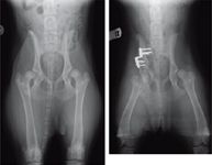
Examination findings include: conformational abnormalities – squared-off hind quarters due to dorso laterally displaced greater trochanters, hind-limb atrophy and laxity and subluxation of the hip, which may be elicited in the relaxed-dog (Ortolani) test. Radiographs will show mild to severe subluxation of the hips (Figure1).
Remodeling changes may appear before 1 year of age in severely affected dogs. There is a wide range of clinical presentations in both the severity of the lesions and severity of clinical signs, yet there is not necessarily a correlation between the severity of the radio graphic lesions and severity of clinical signs.
As the dog matures, clinical signs usually lessen in severity, often by 16 months to 18 months of age. The coxo femoral instability produces hypertrophy of the joint capsule, laying down fibrous tissue and remodeling of the joint that has the effect of stabilizing the joint and reducing lameness. A patient may remain relatively asymptomatic for several years, even in some cases for the rest of its life. But the degenerative processes present in the joint continue as the dog ages, usually producing a recurrence of lameness in middle age.
The degree of lameness is not necessarily proportional to severity of radiographic signs, however, and some dogs with severe degenerative changes will function fairly well as house pets, while others will be severely affected. Radiographs reveal remodeling and periarticular changes consistent with arthritis.

Figure 1: Radiograph of a 6-month-old Golden Retriever. Note the severe subluxation of both femoral heads., Figure 2: Radiograph of the same dog in Figure 1, six weeks after a right-sided pelvic osteotomy. Note the deeply seated right femoral head compared to the still markedly subluxated left femoral head.
Just as hip dysplasia has a biphasic clinical course, its treatment depends on the phase of the disease and the age of the patient. In immature dogs with mild to moderate signs and radiographic changes, conservative therapy can be successful (moderate exercise, weight control and the judicious use of nonsteroidal anti-inflammatory drugs).

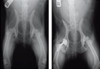
Figure 3: Radiograph of a 7-year-old Labrador Retriever mix. Note the severe remodeling and arthritic changes in both hip joints., Figure 4: Radiographs of the same dog after installation of the cementless total hip. The other hip eventually was replaced, too, giving the animal an excellent return to activity.
Triple pelvic osteotomy
With more severe cases, surgical therapy should be considered. The surgical options that I recommend are the femoral head ostectomy (FHO) and the triple pelvic osteotomy (TPO). In cases where the dog is significantly affected and where remodeling changes are already present, the FHO can be very successful in restoring a functional, pain-free limb, even in larger dogs. The FHO may be selected in cases where there are economic limitations. It should be remembered, however, that the joint is essentially obliterated by the surgery, so the FHO must be considered a salvage procedure.
The goal of the triple pelvic osteotomy is to restore congruency and stability to the hip joint, allowing for normal development of the articular cartilage and prevention of subsequent degenerative joint disease. The procedure, adapted from a surgery performed in children with dysplasia, involves osteotomies of ilial body, ischium and pubis, isolating and freeing the acetabular portion of the pelvis. The acetabulum is rotated to increase dorsal coverage of the femoral head and then secured in that orientation with a special plate that contains a preset angle (usually 20 to 40 degrees). The resultant increased dorsal coverage and tighter fit of the femoral head produces a more stable hip joint (Figure 2).
The short-term objective of the TPO is to produce a stable, less-painful hip. The long-term, and perhaps more important, result of the surgery is to prevent the secondary arthritis seen in the mature dog. When performed in the right patient, the TPO can prevent the ravages of arthritis and restore pain-free hip function in the dog for the rest of its life.
The TPO is reserved for young, moderately to severely dysplastic dogs with no significant remodeling changes visible on radiographs. Most dysplastic dogs will demonstrate remodeling changes by about 1 year of age, and the more severely affected dogs may show remodeling as early as 6 months to 8 months of age. If remodeling is already radiographically evident, then the TPO may only limit arthritic progression. I am reluctant to perform the TPO on dogs older than 1 year of age, or on younger dogs already showing radiographic degenerative changes.
A fairly narrow window of opportunity is present to perform the TPO, so I recommend taking preliminary hip radiographs in a susceptible puppy as soon as a rear-limb lameness or gait disturbance is noted. Radiographs can be taken when the dog is under anesthesia for castration or ovariohysterectomy, but do not delay indicated radiographs if signs are noted prior to a scheduled surgery.
Because both hips normally are affected, I usually recommend that the TPO be performed on both hips. Generally, I will do only one side at a time, with an interval of two to six weeks between surgeries. Postoperatively, dogs will remain in the hospital for two to three days and strict activity restriction is recommended for six weeks following surgery. Although expensive, the results of the TPO surgery can be extremely gratifying to the owner, and spare the dysplastic dog a lifetime limited by pain.
Total hip replacement
Treatment for mature dogs who are less severely affected also can be conservative, and many dogs can be successfully managed this way for the remainder of their lives. In more severely affected individuals, a femoral head excision can again be employed with great success.
But in larger dogs, total hip replacement (THR) surgery can be considered. THR surgery, where the arthritic femoral head and acetabulum are replaced with plastic and metal components, is commonly performed in humans with severe arthritis of the hip joint and has become much more available and common for dogs in the last two decades (Figure 3).
Until the last few years, the standard technique for THR in dogs secured the hip implants to the bone with cement. While this is very strong initially, the implants can loosen with time, and revision surgery or even implant removal may be required. Although good to excellent results usually are obtained, the cemented system may, in a small number of cases, contribute to complications such as infection.
A cementless hip replacement system recently became available as well. The femoral stem and acetabular cup have specially prepared surfaces that allow for ingrowth of bone, providing long-term stability without cement (Figure 4).
In most cases, this fixation will be more secure, last longer and lessen the chance of loosening and other complications. Although the cementless implants require precise preparation of the parent bone, the fact that there are no cement or screws to secure the implants allows for decreased surgical time. In some cases, a combination of cemented and cementless implants can be used to achieve optimum results.
Podcast CE: A Surgeon’s Perspective on Current Trends for the Management of Osteoarthritis, Part 1
May 17th 2024David L. Dycus, DVM, MS, CCRP, DACVS joins Adam Christman, DVM, MBA, to discuss a proactive approach to the diagnosis of osteoarthritis and the best tools for general practice.
Listen