Cholangiohepatitis in race horses
Caring for those affected by the disease that caused champion Uncle Mo to be scratched from the Derby.
The 2010 Breeders' Cup and 2010 Juvenile Champion, Uncle Mo, came into his 3-year-old season the overwhelming favorite for the 2011 Kentucky Derby. But several weeks before he was set to run, he became ill, and only two days before the race, May 5, 2011, he was scratched.

(WILLIAM R. SALLAZ/GETTY IMAGES)
The illness took the potential Triple Crown contender to the sidelines for extensive testing. After several weeks, Bill Bernard, DVM, Lexington, Ky.; Doug Byars, DVM, Dipl. AVCIM, Dipl. AVECC, Byars Equine Advisory, Georgetown, Ky.; and Tom Divers, DVM, professor of clinical sciences, Cornell University College of Veterinary Medicine, diagnosed cholangiohepatitis, an inflammation of the bile passages and liver, based on a biopsy of his liver and nearby lymph nodes. In a statement issued June 3, 2011, the internists reported that they did not know how he contracted the disease.
"Uncle Mo was placed on medications prior to the Derby, though he did not get antibiotics or a liver biopsy prior to scratching from the race," says Byars. "We just didn't have enough time for this horse to recover before the race."
Etiology
According to The Merck Veterinary Manual, the etiology of cholangiohepatitis is likely related to bacteremia that is due to an organism eliminated in the bile, an ascending infection of the biliary tract after an intestinal disturbance or ileus.1 Horses may have an initiating ascending gram-negative infection, but gram-positive aerobic or anaerobic infection is possible, too.
If present, the ascending infection is "thought to initiate a cholangitis, which may extend to the periportal region of the liver," says Divers.
Jennifer Davis, DVM, PhD, assistant professor of equine internal medicine and clinical pharmacology at North Carolina State University College of Veterinary Medicine, reports that although bacteria via ascending proximal small intestinal infection or inflammation may occur, bacteria or bacterial products (such as endotoxins) or inflammatory mediators in the portal circulation might also cause periportal neutrophilic inflammation.
However, not all cases are caused by bacterial infection. "There are many causes of cholangiohepatitis—bacterial, viral, toxic and immunologic," says Divers.
Davis describes two possible pathogenic mechanisms: The first is due to an ascending infection from the proximal small intestine characterized by acute inflammation of the wall of the bile ducts (cholangitis) and entry of large numbers of neutrophils directly into the lumen of the biliary ducts. The inflammatory response can extend into the periportal hepatic parenchyma, causing cholangiohepatitis. Fluid accumulation in the small intestine secondary to ileus and increased luminal pressure could increase the chances of an ascending infection by forcing bacteria into the bile duct. Increased luminal pressure may also reduce bile flow.
The second mechanism involves induction of neutrophilic infiltration by bacteria, bacterial products, inflammatory mediators or a combination of causes in the portal circulation. "Injury to the intestine from a variety of causes can increase the permeability of the mucosal barrier," says Davis. "Absorption of bacteria or their products [particularly endotoxins] promotes intestinal inflammation."
The inflammatory response in the intestine might be associated with the appearance of mediators such as interleukin-8, tumor necrosis factor alpha and interleukin-1-beta in the portal blood. "The effects of these mediators on endothelial cells and neutrophils in portal circulation could induce periportal inflammation and, eventually, suppurative cholangiohepatitis," Davis says.
Clinical signs
The clinical signs of cholangiohepatitis vary depending on the severity of infection and the organism involved, and the signs may be acute, subacute or chronic.1 Anorexia, weight loss, intermittent or persistent fever or colic are often associated with subacute or chronic cases. For these horses, icterus, increased bilirubin and total bile acid concentrations as well as elevated liver enzyme activities are also common. A swollen, soft, pale liver is indicative of acute cholangiohepatitis.
Davis notes that in severe cases, other findings include signs of hepatic failure such as hepatic encephalopathy, colic with associated increases in hepatic enzyme activity, histopathologic evidence of cholangiohepatitis associated with large colon displacements and ulcerative duodenitis in foals and yearling horses. "This suggests GI disease might be a predisposing factor," says Davis.
In addition to cholangiohepatitis, some horses also have cholelithiasis. "Horses get stones in their livers sometimes associated with infections or from a dietary association—soft brown stones, and they're usually crushable," says Byars. "If they end up going to surgery, the stones are crushed, so the surgeon doesn't have to remove them."
Horses don't have gallbladders, so when those channels are affected (i.e., plugged), they don't have residual bile storage.
Diagnosis
"A lot of these horses are not jaundiced; many are functioning reasonably well," says Byars. "Most are not at a crisis, except for a small percentage that Dr. Peek and Dr. Divers describe as being end-stage. It's kind of like a person who has the blahs—doesn't feel consistently well."
The liver is the body's processing plant. "So, if the liver is having difficulties, energy level and the detoxification process are being slowed," Byars says. "Metabolically, that would be compromising and can lead to varying clinical signs. Horses are also unique in that they can be off feed for some other reason that has nothing to do with liver disease and develop icterus called anorectic icterus. Their bilirubin will increase—not terribly, but way out of the normal range. So, for some of those horses that just don't eat, they go back on feed, and then it goes away.
"The bottom line is to pull lab work," Byars says. "If you have a horse that's not doing well, you probably don't suspect hepatitis or cholangiohepatitis. Within the blood work there are certain liver enzymes that are not only produced by the liver but also are cleared by it. And there are some enzymes that are not produced in particularly great amounts by the liver but are also cleared by it."
Gamma-glutamyltransferase (GGT) can be a marker found in horses that reflects suspicion of liver involvement, according to Byars. Several published reports have found increased GGT activity in performances horses that have decreased performance without apparent reason. "Many of those don't perform well, but they get better," Byars says. "Whether they had a cholangiohepatitis or something similar, you'd never know unless the enzyme elevations continue to produce a predictable pattern or you do a liver biopsy. Obviously, horses in training aren't candidates for liver biopsy."
The enzymes alkaline phosphatase (ALP) and GGT are cleared through the liver; GGT is also produced by the liver. "When those two simultaneously increase, you begin to think of reduced bile flow," says Byars.
Liver enzymes such as lactate dehydrogenase (LDH) and aspartate transaminase (AST) are found in many parts of the body and are, therefore, not liver-specific. "If you have elevated GGT activity by itself, you may suspect a clearance problem. If you also have increased ALP, then you know the bile flow is reduced and most likely you have a cholangitis, and possibly cholelithiasis."
Diagnostics should include biochemical tests (SDH, AST, LDH, ALP, GGT, serum bile acids, serum or plasma bilirubin) to determine whether there's liver involvement.

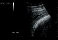
Photo 1: An ultrasonogram of the liver next to the spleen in a horse with cholangiohepatitis. Sludge can be seen in the bile duct. (Image courtesy of Dr. T.J. Divers, Cornell University.)
Transabdominal ultrasonography is another tool that can help diagnose cholangiohepatitis (Photos 1-3). While ultrasound-guided liver biopsy is required to make a definitive diagnosis, repeat ultrasonographic evaluations of the liver can also be useful in assessing the success of therapy.

Photo 2: An ultrasonogram of a distended bile duct in a horse with cholangiohepatitis. (Image courtesy of Dr. T.J. Divers, Cornell University.)
"Ultrasound can reveal the presence of abscessation or neoplasia, dilation of bile ducts, presence of stones and an assessment of the hepatic texture or changes in the liver lining consistent with nodular formation," says Byars.

Photo 3: An ultrasonogram of a stone in the bile duct casting a shadow in a horse with cholangiohepatitis. (Image courtesy of Dr. T.J. Divers, Cornell University.)
Ultrasonographically, cholangiohepatitis may appear with both dilated bile ducts, reflecting reduced bile flow, and textural changes to the hepatic parenchyma. However, the findings may be highly variable in many horses that appear relatively normal but have abnormal chemistry results, according to Byars.
"Many veterinarians will skip a full chemistry, maybe just run a CBC, looking for infection, etc.," says Byars. "Practitioners should take the clinical complaints, combine those with a complete blood chemistry panel and use ultrasound. A biopsy would be definitive. If you have 100 suspected liver-involved horses in the field, maybe 10 percent get biopsied, but those at referral hospitals typically are biopsied."
Recognizing certain liver enzymes that are failing to be cleared appropriately in conjunction with increased liver-specific enzymes (GGT and AP) would help to support a diagnosis of cholangiohepatitis.
Treatment
Treatment for cholangiohepatitis is to change the diet, Byars says. "You don't want too high a protein content. Feeding a very digestible feed such as Equine Senior or a beet pulp-based diet is suggested, along with fat for energy and vitamin supplementation."
Such supplementation should be mostly B vitamins. Avoid excessive fat-soluble vitamins such as vitamin A, because they may lead to toxicosis if the liver isn't metabolizing it properly. "Vitamin E is OK, but you wouldn't supplement with vitamin D," Byars advises. A B-complex vitamin supplement along with vitamin E as an antioxidant would be ideal, he says.
Antibiotics may also be indicated. "An antibiotic that goes through the enterohepatic circulation cycle is helpful—one that is taken orally, absorbed, goes through the liver, goes back to the gut and back to the liver," says Byars. "It cycles through the liver and bathes that system to treat an infection. Potentiated sulfa tablets such as trimethoprim-sulfadiazine are the best known to do that.
"You put horses on an antibiotic and subjectively treat a bacterial infection. The only way it would not be subjective would be if you got a confirmation culture from the biopsy and isolated an organism," says Byars. "But you're usually not able to do that and subjectively treat and see how they respond. Pentoxifylline is an immunoregulatory agent that has been documented in many species, including humans, to be safe for healing the liver, blocking some of the end-stage inflammatory fibrosis, tumor-necrosis factor and some of the cytokines.
"Another drug, colchicine, is anti-inflammatory and is subjectively used, sometimes in conjunction with pentoxifylline," Byars says. "Ursodiol, a bile acid, may be administered to change bile acid content and reduce cholelithiasis."
Prognosis
Cholangiohepatitis can advance to hepatic failure, but treatment is often successful, and the prognosis usually is favorable, says Byars. "It takes some time to heal."
Practitioners should carefully monitor the animal. "Liver enzymes give you a good idea of what the progress or healing is, as does the horse's clinical status," Byars says. "If you biopsied the animal and there was significant fibrosis, you're in trouble. If there was a lot of bridging of the hepatocytes with fibrous deposition, you would know you were pretty close to an end-stage case, which would be difficult to turn around due to so much scarring."
Increased liver enzyme activity in these horses with advanced disease may be minimal since the cells that produce them are no longer functioning. "Clinically, end-stage liver horses will be thin, partially anorectic and have behavioral abnormalities," says Byars.
The time course for healing varies with each individual and disease severity. The liver is a regenerative organ. "Remove the insult, treat them symptomatically, and many of these horses will respond successfully to treatment within a month to a year. Most cases will improve within three months," says Byars. Often, performance horses treated early will be well on their way to recovery with simple dietary changes and minimal medication.
As for Uncle Mo, it was reported on June 8 that his health was improving nicely, and it was hoped that he would resume training by late June.
Ed Kane, PhD, is a researcher and consultant in animal nutrition. He is an author and editor on nutrition, physiology and veterinary medicine with a background in horses, pets and livestock. Kane is based in Seattle.
Reference
1. Kahn CM, Line S. Merck Veterinary Manual. 10th ed. Whitehouse Station, NJ: Merck & Co, 2010.
Suggested Reading
- Davis JL, Jones SL. Suppurative cholangiohepatitis and enteritis in adult horses. J Vet Intern Med 2003;17(4):583-587.
- Peek SF, Divers TJ. Medical treatment of cholangiohepatitis and cholelithiasis in mature horses: 9 cases (1991-1998). Equine Vet J 2000;32(4):301-306.
Podcast CE: A Surgeon’s Perspective on Current Trends for the Management of Osteoarthritis, Part 1
May 17th 2024David L. Dycus, DVM, MS, CCRP, DACVS joins Adam Christman, DVM, MBA, to discuss a proactive approach to the diagnosis of osteoarthritis and the best tools for general practice.
Listen