Dental and oral examination: A visual atlas of dental and oral pathology (Proceedings)
The many abnormalities, lesions and diseases that are commonly found in the oral cavity become much easier to recognize after one has become familiar with the normal oral anatomy and structures. The most common and most obvious problems, such as periodontal disease and fractured teeth, are easy to identify during routine oral examinations.
The many abnormalities, lesions and diseases that are commonly found in the oral cavity become much easier to recognize after one has become familiar with the normal oral anatomy and structures. The most common and most obvious problems, such as periodontal disease and fractured teeth, are easy to identify during routine oral examinations. But there are a variety of other diseases and lesions that routinely occur. Some of the things that can be visually identified include swellings (tumors, endodontic abscesses, developmental cysts, trauma, hyerplasia), tooth resorption, caudal stomatitis, super-eruption (most commonly canine teeth in cats), developmental anomalies, embedded or impacted teeth, hypodontia, supernumerary teeth, dental crowding, malocclusion/traumatic occlusion, and systemic diseases with oral symptoms.
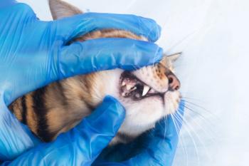
Dogs and cats can suffer from most of the same dental problems from which humans suffer, plus some additional ones peculiar to their anatomy and habits. Pets are often stoic, showing no outward signs even when there is significant discomfort. For this reason it is important to lift the lips and open the mouth, and to perform a thorough oral examination of every patient that is evaluated – regardless of the reason for the scheduled general physical exam. The oral cavity is a very important structure with an interesting and complex anatomy and micro-environment. A healthy mouth is needed for optimum nutritional intake, comfort, and over-all health.
This session is designed as an overview to show the clinical appearance of a variety of dental and oral pathologies. Seeing the clinical appearance of lesions makes them easier to recognize when they are seen again in your patients.
Periodontal disease
This is the most common problem that we will see in our patients. Bacterial plaque is a biofilm that lives on the surfaces of the teeth as well as other oral surfaces such as the dorsum of the tongue. This biofilm is antigenic and causes gingivitis where the gingiva contacts it at the gingival margin. Gingivitis is diagnosed by clinically observing red (mild), edematous (moderate), or ulcerated (severe) gingiva. After only a few days on a tooth surface it will mineralize into calculus ("tartar"), a hard tightly adherent material. Subgingival plaque along with the immune response of a susceptible host will cause bone loss and destruction of the attachment of the tooth. This is periodontitis and is identified clinically as gingival recession, deep periodontal pockets, tooth mobility, or bone loss on a radiograph.
Stomatitis
Any tissue in the mouth can be inflamed; mucositis, glossitis, palatitis, cheilitis, etc. Generalized inflammation of the mouth, or stomatitis, can be frustrating to treat because it is often difficult to identify the cause. The most common cause of generalized gingivitis is plaque on the tooth surfaces. When this inflammation extends to the mucosa it is no longer a normal inflammatory response. A common form of this is chronic ulcerative stomatitis in which mucosal ulceration is present at multiple sites. The most commonly affected sites are where the mucosa of the cheeks or lips contacts large tooth surfaces such as the canine tooth and the maxillary fourth premolar. These focal mucosal ulcerations are called contact ulcers. Severe inflammation in these areas often results in gingival recession. Some of the causes can include metabolic disease (renal failure with generalized uremic stomatitis), autoimmune diseases such as pemphigus vulgaris, and eosinophilic stomatitis. Some breeds, including Greyhounds and other sight hounds, are predisposed to stomatitis due to an incompletely understood immunopathy.
Feline caudal stomatitis
A specific form of stomatitis that occurs in the caudal tissues of cats that spans the space between the retromolar area of the upper and lower jaws. This is the region of the pterygomandibular raphe, and inflammation often first affects the palatoglossal folds. In severe cases it will extend down the throat to the fauces and further, and laterally to the cheeks. Clinical observation of (sometimes severely) ulcerated and proliferative tissue bilaterally in these areas is characteristic of this syndrome.
Trauma
Trauma to the teeth, oro-facial bones, and soft tissues is a common finding but can sometimes be difficult to identify. Mandibular fractures and temporomandibular joint dislocations cause asymmetry with the mandible displaced towards the side of a fractured mandible or away from the side of TMJ dislocation. Fractured teeth are easy to diagnose with a quick observation, but even these can be hidden under calculus accumulations or on the lingual side of teeth. Treatment of traumatized teeth is covered in another session.
Maxillary bony trauma usually results in minimal displacement of fragments even with severe trauma. Whenever a patient has suffered trauma, look for palatal and mandibular integrity, mandibular excursions to rule out temporomandibular joint pathology, mobile or malpositioned teeth, and soft tissue tears including on the tongue.
Tumors and cysts
Tumors can appear as discrete enlargements affecting any oral tissues. They can also manifest as diffuse enlargements of facial tissues, areas of normal architecture but abnormal texture or firmness, deviated teeth, one or more mobile teeth in a single region in the absence of periodontitis, deviation of the tongue, tonsillar enlargement, pigmentary changes, or pathological fracture. The most common tumor in the dog is the peripheral odontogenic fibroma (previously characterized as fibromatous or ossifying epulis). This is a benign tumor that is very firm, non-ulcerated and involving the subjacent bone. Complete removal requires including soft tissue and bony margins down to the involved tooth roots including the periodontal ligament and cementum. This usually requires removal of the contact teeth as well in an en bloc resection.
Squamous cell carcinoma is the most common neoplasia in cats. It is often diagnosed late when local surgical control can be challenging.
There are many oral and dentigerous cysts that occur. One of the most common is a dentigerous cyst associated with an unerupted mandibular (and less commonly maxillary) first premolar tooth. These enlarge by expansion and can become dramatically large as they displace the mandibular bone.
Most inflammatory cysts are related to endodontic disease.
Compound odontomas and complex odontomas are an infrequent cause of swelling. Radiographs help differentiate them from other masses.
Deep infection
Osteomyelitis can be caused by extension of endodontic or periodontal infection or from penetrating wounds. The clinically apparent sign is swelling that can be either visualized or palpated. Radiographs and sometimes biopsy are necessary for diagnosis.
Orbital cellulitis
Infection from penetration of a foreign body into the tissues behind the maxillary second molar, or from extension of dental infection of the first or second molar can result in orbital cellulitis or retrobulbar abscess. The most common and obvious symptom is extreme pain on opening the mouth. Other symptoms include proptosis and inability to retropulse the globe on the affected side, nictitans prolapse and scleral injection.
Tooth resorption
There are many known causes of tooth resorption (TR) including inflammation, physiologic, pressure etc. However there is also an ideopathic tooth resorption that is very common in cats and also occurs in many other species, although much less frequently, including dogs and humans. This TR is usually first identified close to the gingival margin of the tooth. Patients often have multiple teeth affected, so full mouth radiographs are recommended for affected individuals. A sharp-edged defect that is often filled with granulation tissue on the surface of the tooth close to the gingiva is the distinguishing feature. In cats the first tooth affected is often the mandibular third premolar (the first tooth behind the lower canine) and in dogs it is often the mandibular first molar (carnassial).
Super-eruption
Periodontal disease around the canine teeth of cats can result in an expansile alveolitis in which the bone around the root appears enlarged. It is actually lifted away from the root. These teeth often super-erupt as inflammation around the root tip slowly pushes the tooth from the socket. This gives the appearance of a very long canine tooth. It can affect one or all of the canines. The cementoenamel junction can be seen a few millimeters coronal to the gingival margin, but it is due to the coronal movement of the tooth rather than apical movement of the gingiva (gingival recession). Affected teeth are typically very firmly attached with little-or-no mobility, even when only a small amount of apical tooth is all that remains supported by bone.
Hypodontia and polyodontia
Supernumerary or congenitally missing teeth are common. When an extra tooth causes crowding or inflammation it should be removed. Supernumerary premolars in dogs often cause no problem since in many breeds there is enough space to accommodate them. In small breeds or cats, supernumerary teeth frequently must be removed. Congenitally missing teeth are rarely a serious problem other than for owners that had aspirations for show careers for their dog or cat.
Embedded teeth
Clinically missing teeth may have been congenitally absent, exfoliated from disease, fractured subgingivally, or they could also be present but unerupted. A radiograph should be made to determine which of these etiologies was responsible since all but the first could require intervention.
Crowding
The most common cause for crowding is genetic manipulation intending to shorten the face to get a blockier head shape or outright brachycephaly. The jaws tend to decrease in size more than the teeth do, resulting in crowding that usually predisposes to periodontal disease. The most commonly affected site is the maxillary premolar area.
Crowding can also occur in areas of supernumerary teeth if there is insufficient space to accommodate the extra teeth. Cats with extra premolars rarely have enough space for them without resulting in significant periodontal disease or occlusal interference.
Dental anomalies
Developmental anomalies of the teeth and jaws can be genetic, either random or by intentional or unintentional design. Another cause of developmental anomalies is injury to the tooth buds or the tissues that surround them during tooth development. Trauma-induced anomalies can affect a single tooth or a number of teeth in one area. Some anomalies are of interest but of no clinical significance. Others can run the entire range of minimal to severe impact on health, many requiring intervention. Some of the anomalies that can be seen (besides polyodontia and hypodontia described above) include fused teeth, dental bigeminy, dens invaginatus (dens-in-dente), dens evaginatus, dental twinning, dilacerations of the crown or of the root, interrupted development of the enamel (hypoplasia or hypomaturation) or of the roots (radicular dysgenesis), dilaceration (severe change in angulation), delayed eruption, embedded and impacted teeth, peg teeth, rotated teeth, malpositioned teeth and malocclusions, compound odontoma, various types of cleft palate, microglossia, etc.
Summary
Dental and oral disease can have a profound negative effect on the health of our patients by causing discomfort, inflammation, infection, loss of function, and premature death. Many of the pathological processes of the teeth and oral cavity remain hidden and apparently asymptomatic until late in their course, but they can be discovered early with proper oral examinations. Dental and oral examination should be an important part of every physical examination done on our patients. Knowing what to look for during these oral exams helps us to make accurate diagnoses.
Newsletter
From exam room tips to practice management insights, get trusted veterinary news delivered straight to your inbox—subscribe to dvm360.


