Dorsal cervical articular process injection: when and how (Proceedings)
Osteoarthritis and cervical pain can present with clinical. Almost all skeletal preparations of horses over 18 years old have evidence of arthritic changes.
Osteoarthritis and cervical pain can present with clinical. Almost all skeletal preparations of horses over 18 years old have evidence of arthritic changes. The disease occurs with some frequency in horses at competitive event ages. Osteoarthritis and other degenerative changes initiated via acute or chronic trauma, or osteochondrosis of articular process articulations are common etiologies.
Ultrasonographic anatomy
Figure 1 is a skeletal preparation of C4 and C5 and depicts the gross anatomy of the structures in question. The area of synovial attachment has evidence of osteophyte formation (c). The caudal C4 facet (a) cranial C5 facet (b) and transverse process of C4 (d) are evident and labeled.
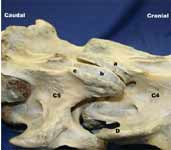
Figure 1: A right lateral view of a skeletal preparation of cervical vertebrae C4 and C5. See text for legend.
The cervical articular facets form an identifiable ultrasonographic landmark that has the appearance of a hand with an outstretched thumb and flexed 2 first digits (Figure 2).

Figure 2: A bone preparation and ultrasound image of the right vertebral articulation of C4 and C5. See text for legend.
In Figure 2, the image of the hand (right) represents the ultrasonographic image. The caudal facet of C4 (C) and its transverse process (E), and the cranial facet of C5 (B) and its transverse process (D) are labeled. The ultrasound probe (A) is at the approximate scanning position as the image in the center. The ultrasound image in the center shows the bone shadows of the transverse process of C4 (E), the cranial facet of C5 (B) and caudal facet of C4 (C). The image of the hand is likewise labeled with the caudal facet of C4 (C) and its transverse process (E), and the cranial facet of C5 (B).
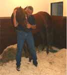
Figure 3: A method of evaluating neck flexibility.
Identifying the site for anti-inflammatory drug injection
Physical examination specific for neck pain
Physical examination of the horses with cervical neck pain indicates reduced to poor cervical flexibility. This author considers normal flexion evident when the horse's nose can be flexed to the left pant pocket with smooth motion and little hesitation. Some training may be necessary. These procedures should be performed carefully, consistently and bilaterally.
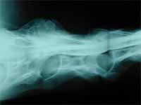
Figure 4: Cervical myelograph with evidence of severe degenerative changes of the right dorsal vertebral articulation of C6 to C7.
Radiography
Radiographic changes with little to no clinical signs have been seen by the author. Clinical However, severe pathology must be present for radiographs to be diagnostic (Figure 4, 5, and 6).
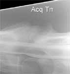
Figure 5: Cervical radiograph that shows severe degenerative changes of the dorsal vertebral articulation of the C5-C6 articulation.
Ultrasonography
Ultrasonographically, the soft tissues including the synovial joint space and synovial attachments are visible and abnormalities are clearly identified (Figure 7).
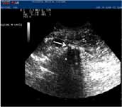
Figure 6: Ultrasonographic image of dorsal lateral cervical articulation with evidence of osteophyte formation (arrow). The caudal C5 facet (A) is evident.
Nuclear scintigraphic imaging
Nuclear scintigraphic imaging can also be helpful for identifying problematic articulations (Figure 8).

Figure 7. Ultrasonographic image of dorsal lateral cervical articulation from 2 horses. In the left image, there is evidence of severe disruptive disease as compared to the normal image on the right.
In Figure 8, the image to the left is considered a normal cervical scintigraphic image. The center image has evidence of increased uptake at the C6 and C7 articulation (arrow) which indicates extensive bone remodeling likely due to inflammation. The image at the right indicates increased ventral articular uptake at C4 and C5 (arrow). Arguably all the caudal dorsal articulations have increased uptake as well.

Figure 8: Scintigraphic images of 3 horses (Courtesy of Dr. Tina Kemper and San Luis Rey Equine Hospital). See text for legend.
Methods
The procedure will be discussed during the presentation.
The arrow in the left image in Figure 9 shows the caudal facet of C6 and points to the capsular attachment.
A dose of 40 to 120 mg methylprednisolone or up to 30 mg of celestone are used by the author. Amikacin at 125 mg can be added, but the author feels this is unnecessary. Volumes of more than 3 to 4 ml should be avoided per articulation. The technique can be repeated at additional sites as needed.

Figure 9: Two images of a gross anatomical preparation of cervical vertebrae 6. See text for legend.
References and additional reading
Denoix J-M, D. S. (2003). Thoracolumbar spine. In M. Ross, & S. Dyson (Eds.), Diagnosis and management of lameness in the horse (pp. 509-521). Philadelphia: WB Saunders.
Grisel GR, G. B. (1996). Arthrocentesis of the equine cervical facets. Preceedings of the AAEP, 42nd Annual Convention, (pp. 197-198). Denver.
Mattoon JS, D. W. (2004). Technique for equine cervical articular process joint injection. Vet Radiol Ultrasound, 45 (3), 238–240.
Podcast CE: A Surgeon’s Perspective on Current Trends for the Management of Osteoarthritis, Part 1
May 17th 2024David L. Dycus, DVM, MS, CCRP, DACVS joins Adam Christman, DVM, MBA, to discuss a proactive approach to the diagnosis of osteoarthritis and the best tools for general practice.
Listen