Equine Image Quiz: Neurologic deficits and vestibular dysfunction in a palamino
Correct answer for Equine Image Quiz: Neurologic deficits and vestibular dysfunction in a palamino
Temporohyoid osteoarthropathy is correct!
This disorder should be the primary diagnostic differential in cases of acute onset of peripheral vestibular dysfunction with facial nerve paralysis.
Infectious, traumatic, and degenerative conditions are potential causes for the development of temporohyoid osteoarthropathy. Neurologic dysfunction is an acute manifestation of chronic bony proliferation of the petrous temporal and stylohyoid bones, resulting in ankylosis of the temporohyoid joint and secondary fracture of the petrous temporal bone. A fracture line extending into the cranial vault at the level of the internal auditory meatus will induce direct trauma to the vestibulocochlear and facial nerves with associated clinical signs of vestibular dysfunction and facial nerve paralysis. If the fracture line extends to the foramen lacerum, caudal to the petrous temporal bone, trauma to the glossopharyngeal and vagal nerves can occur, resulting in dysphagia. In addition, inflammation from the disease process may extend through the internal acoustic meatus and lead to focal suppurative meningitis at the level of the pons, resulting in fever and depression.
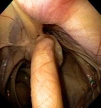
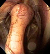
Endoscopic examination of the right guttural pouch showed osseous proliferation of the proximal insertion of the stylohyoid bone at the level of the temporohyoid joint. Note the marked enlargement of the right stylohyoid bone in comparison to the left.


Left guttural pouch (left) vs. right guttural pouch (right)
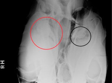
A standing ventrodorsal radiograph revealed sclerosis of the tympanic bulla and proximal portion of the stylohyoid bone. Note the bulbous enlargement of the proximal portion of the stylohyoid bone at the base of the petrous temporal bone on the right side (red circle) compared with the left side (black circle). The actual fracture of the petrous temporal bone could not be clearly identified in this case, probably because of minimal displacement of the fragments.

A recovery rate of up to 50% has been reported after long-term medical treatment with broad-spectrum antimicrobials, anti-inflammatory drugs as well as analgesic and corneal ulcer therapy as needed. Surgery is also an alternative treatment option and can be performed before or after the development of neurologic deficits. A partial stylohyoid ostectomy (the removal of a 2-cm midshaft segment of the stylohyoid bone) can be performed to decrease the tension on the temporohyoid joint and the risk of fracture of the petrous temporal bone. Alternatively, a ceratohyoidectomy (disarticulation of the ceratohyoid bone from the basihyoid bone, followed by disarticulation of the ceratohyoid-stylohyoid joint) can be done on the affected side.
In this case, surgery was declined and medical management was provided. Only subtle head tilt and mild facial nerve paralysis persisted after a six-week course of treatment with oral chloramphenicol and dexamethasone.