Meal-induced hyperadrenocorticism in dogs: Not to be overlooked
When to suspect that food is at fault-and what to do about it.
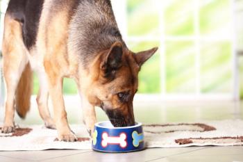
Adrenal glands, meet your enemy. (Getty Images)
Cushing's disease in dogs can be diagnostically frustrating to veterinarians in clinical practice. As clinicians, we frequently suspect the problem and spend our time and clients' money desperately trying to prove a diagnosis, or we find ourselves chasing laboratory abnormalities with yet more laboratory testing.
David Bruyette, DVM, DACVIM, medical director of VCA West Los Angeles Animal Hospital, defines Cushing's syndrome as “basically encompassing any form of adrenal overproduction of steroids.” Five to ten percent of dogs with classic clinical signs of hyperadrenocorticism will have negative or equivocal test results on standard laboratory tests including the ACTH stimulation test, low-dose dexamethasone suppression (LDDS) test and urine cortisol:creatinine ratio (UCCR), says Bruyette.
At the 2015 CVC San Diego this December, Bruyette presented a detailed overview of both classic Cushing's disease and various types of “other syndromes that either result in an overproduction of cortisol or an overproduction of sex steroids.” One of these lesser known forms of Cushing's disease, called meal or food-induced Cushing's disease, may be important to have on our differential lists as clinicians.1
Not your average everyday Cushing's
“Regular old boring Cushing's disease,” as Bruyette fondly coins it, is the result of a benign or malignant ACTH-secreting or glucocorticoid-secreting tumor in the pituitary gland or adrenal glands, respectively. Iatrogenic disease or exogenous sources of cortisol such as corticosteroid-containing cream are other causes of Cushing's syndrome that, while sometimes confounding diagnostically, have more obvious treatment options and are not as pathophysiologically confusing as the endogenous forms.
The remaining nonclassic, less well-understood categories of Cushing's syndrome etiologies include ectopic ACTH production by a tumor, meal- or food-induced Cushing's disease, cyclical hyperadrenocorticism, occult hyperadrenocorticism and atypical hyperadrenocorticism, says Bruyette. Some of these syndromes produce clinical signs and laboratory abnormalities on routine serum chemistry profiles and adrenocortical function tests, and some do not.
Patients with an overproduction of adrenal steroids-including cortisol and adrenal sex steroids-that have a lack of or mild serum chemistry profile abnormalities (e.g. mildly elevated phosphate concentration and alanine transaminase activity, equivocal adrenal function test results, or merely elevated sex steroids on an adrenal steroid profile from the University of Tennessee Medical Center Laboratory) but that demonstrate no clinical signs or just an endocrine alopecia are less urgent and less clinically relevant. They may not need treatment at all. From a clinical perspective, all forms are most important when they cause classic clinical signs of Cushing's disease-polyuria, polydipsia, hair coat changes and a pendulous abdomen. In addition to the deleterious long-term effects of cortisol on the entire body, these clinical signs are distressing to clients, and they will come to us for answers and a resolution to the problem.
A clue to a cause from human medicine
In humans with meal- or food-induced hypercortisolemia, patients are born with a congenital defect that results in the aberrant expression of receptors for glucagon inhibitory peptide (GIP) on the adrenal gland.2 When stimulated by GIP, the adrenal glands in these patients produce cortisol. GIP is produced by the stomach during every meal, and it normally binds to receptors on the pancreas to stimulate insulin production. In a food-induced hyperadrenocorticism patient, the GIP released at every meal abnormally triggers the release of cortisol by binding to these aberrant receptors on the adrenal gland. This appears to be the case in dogs as well.
Bruyette says, “If you look at the adrenal glands of these dogs either grossly or histologically, you can see that they have a lot of these GIP receptors being expressed all throughout their adrenal glands.” Therefore, these dogs have transient hypercortisolemia during every meal from a young age. When they present to our clinics many hours after a meal or occasionally fasted for adrenocortical function testing, the test results are negative because “it's not a constant state of hypercortisolism, it's an intermittent state of hypercortisolism,” Bruyette says.
Which dogs are at risk?
According to Bruyette, dogs in which meal- or food-induced hyperadrenocorticism is diagnosed are young, with an early onset of clinical signs between 2 and 5 years of age. Bruyette and other clinicians have found no breed predilection, but so far he says all of the cases have been in small dogs. “These are dogs that look floridly Cushingoid,” says Bruyette. They have classic clinical signs of hypercortisolemia, including polyuria, polydipsia, polyphagia, muscle wasting, a pendulous abdomen, hepatomegaly with a vacuolar hepatopathy, and dermatologic changes such as truncal alopecia or even calcinosis cutis.
Laboratory abnormalities such as elevated alkaline phosphatase activity are present, and the adrenal glands are usually bilaterally enlarged on abdominal ultrasonographic examination, but the ACTH stimulation test, LDDS test, and UCCR results are all normal.1 If the diagnosis is not properly made in a timely manner, these dogs face many more years of progressively worsening disease than the average geriatric Cushing's patient.
The nitty-gritty on diagnosis and treatment
Food-induced hyperadrenocorticism is fairly easy to diagnose and relatively simple to treat, as long as you know when to suspect it and which diagnostic tests to choose. Although the incidence of this problem appears to be low, any untreated form of chronic hypercortisolism has high morbidity, so as clinicians we should try not to miss these dogs with food- or meal-induced Cushing's disease.
The basic diagnostic work-up for Cushing's disease should have already been completed-a complete blood count, a serum chemistry profile, a urinalysis, an abdominal ultrasonographic examination, plus “ideally at least two adrenal-pituitary function tests,” Bruyette says, such as an LDDS test, an ACTH stimulation test or a UCCR.
If you suspect meal-induced hyperadrenocorticism in a dog, at this point “it's a very simple thing to diagnose,” says Bruyette. Have the clients fast the dog for 12 hours and obtain a urine sample at home. Ideally use the first morning-voided sample. Instruct them to feed the dog and four hours later have them go for a walk and obtain another urine sample. Submit the carefully labeled pre- and post-meal urine samples for a UCCR on each. A 100-fold increase in urine cortisol in response to the meal will be diagnostic for food-induced hyperadrenocorticism. “In general, we do want to at least see the UCCR double, but there are not really enough cases yet reported to show a range,” says Bruyette. “In a normal dog, when he eats, there is no rise in cortisol.”
Treatment for this condition is to give adrenal enzyme blockers two to three hours before every meal. This blocks the effect of GIP on the receptors during GIP stimulation by food, so oral trilostane twice daily with every meal is a very effective treatment. Use the ACTH stimulation testing to monitor treatment as you would to monitor any classic Cushing's disease case. Perform the test four hours after the morning meal, which is about six to seven hours after treatment with trilostane.
References
1. Galac S, Kars VJ, Voorhout G, et al. ACTH-independent hyperadrenocorticism due to food-dependent hypercortisolemia in a dog: a case report. Vet J 2007;177(1):141-143.
2. N'Diaye N, Tremblay J, Hamet P, et al. Adrenocortical overexpression of gastric inhibitory polypeptide receptor underlies food-dependent Cushing's syndrome. J Clin Endocrinol Metab 1998;83(8):2781-2785.
Carla Johnson, DVM, practices emergency medicine at Berkeley Dog and Cat Hospital in Berkeley, California, and general practice at Cameron Veterinary Hospital in Sunnyvale, California. Her nonveterinary loves are writing, Dressage with her Iberian warmblood mare, Synergy; watercolor painting on yupo; vinyasa yoga; and running with her dog Tyson. Try as she might, her curly-coated Scottish Fold, Hootie, refuses to go jogging with her.
Newsletter
From exam room tips to practice management insights, get trusted veterinary news delivered straight to your inbox—subscribe to dvm360.


