Miller's knot case study: step 2
A step-by-step photo package illustrating application of a Miller's knot for veterinarians.
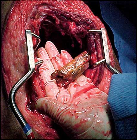
A parasternotomy was completed to allow inspection, drainage and irrigation of both sides of the pleural space as well as removal of the object and, if necessary, lung lobectomy. Upon opening the pleural space it was evident that the stick was lodged deeply into the right cranial lung lobe. The lobe was removed with the accompanying section of stick using the Miller's knot technique.
Here you can see a section of the stick being removed from the right cranial lung lobe. Even after the application of a retaining retractor there is still limited access.
More in this package:
Applying a Miller's knot: step 1
Applying a Miller's knot: step 2
Applying a Miller's knot: step 3
Comparison to traditional technique
