Surgery STAT: Correcting cranial cruciate ligament-deficient stifles
The Tibial Tuberosity Advancement (TTA) is a procedure developed in Zurich, Switzerland, in the early 2000s. The biomechanics behind its success for correcting cranial cruciate ligament-deficient stifles is a change in the patellar tendon angle that effectively neutralizes the tibiofemoral shear (tibial thrust) that occurs during weight bearing.
EDITOR'S NOTE: SurgerySTAT is a collaborative column between the American College of Veterinary Surgeons (ACVS) and DVM Newsmagazine. In November, Lara Rasmussen, DVM, MS, Dipl. ACVS and Stephen H. Levine, DVM, MS, Dipl. ACVS will discuss factors that revolve around their recommendations for medical care for TTA vs. TPLO. In December, Dr. Shawn Mattson, DVM, DVSc, BSc, discusses "Treating subchondral bone cysts in the fetlock joint."
The Tibial Tuberosity Advancement (TTA) is a procedure developed in Zurich, Switzerland, in the early 2000s. The biomechanics behind its success for correcting cranial cruciate ligament-deficient stifles is a change in the patellar tendon angle that effectively neutralizes the tibiofemoral shear (tibial thrust) that occurs during weight bearing.
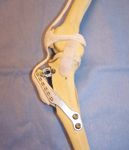
The surgical techniques contributing to its success include a significantly less-invasive osteotomy and the lack of compromise to the weight-bearing axis of the tibia. The procedure involves a cranial tibial crest osteotomy, advancement of the tibial crest with an advancement cage (acting as a stabilizing wedge) and internal fixation of the crest with a thin, tension-band plate held in place with a special fork and bone screws (Image 1).

Image 1: Model replica of Tibial Tuberosity Advancement.
Pre-operative measurements are taken from lateral stifle radiographs (with joint extension) to determine appropriate basket and plate sizes.
In our experience, the patient experience in the immediate post-operative period appears to be improved over other active and passive stabilization techniques such as TPLO and extracapsular lateral suture imbrication. Dogs appear to be more comfortable in the first few post-operative days, as evidenced by early, strong weight bearing. The long-term outcome appears to be equivalent to the TPLO procedure, based on our experience and indirect comparison of published evidence. Patients return to weight bearing rapidly and regain muscle mass quickly. Implant complications are rare, presumably owing to the less-invasive procedure.
Major complications include post-operative patella luxation, tibia fracture, implant loosening and meniscal injury (if initial meniscectomy/meniscal release was not performed). Minor complications include incisional infection/inflammation, seroma and wound dehiscence.
The Tibial Plateau Leveling Osteotomy (TPLO) is a procedure developed in Eugene, Ore., in the early 1990s. It was the first one developed to address the active biomechanics behind cranial cruciate ligament disease. The technique neutralizes tibial thrust during weight bearing by changing the tibial plateau to a neutral angle and effectively recruits the caudal cruciate ligament for primary stability of the stifle.
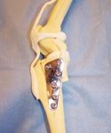
The surgical procedure involves a radial (semi-circular) osteotomy of the proximal tibia, rotation of this proximal tibial segment and internal fixation with a bone plate and screws (Image 2). Pre-operative measurements are taken from lateral stifle radiographs (with partial joint flexion) to determine saw blade and plate size, as well as degree of rotation required.
In our experience, patients recover with better long-term results than traditional passive stabilization techniques, such as extracapsular lateral suture imbrication. By one month post-operatively, patients are strongly weight-bearing and advancing in their at-home physical therapy.

Image 2: Model replica of Tibial Plateau Leveling Osteotomy.
Implant complications, consisting of infections or screw loosening, are seen in a small percentage of cases and are managed with appropriate antibiotics and implant removal after healing.
Major complications include post-operative patella luxation, tibia fracture, implant loosening/failure and implant-related infection, and meniscal injury (if initial meniscectomy/meniscal release was not performed). Minor complications include incisional infection/inflammation, seroma and wound dehiscence.
In the second part of this column, we will briefly discuss the approach to decision-making with regard to these procedures and their application in the clinical setting.
Drs. Rasmussen and Levine are ACVS board-certified surgeons. Dr. Rasmussen is with the Veterinary Surgical Specialists — a small-animal surgery specialty practice serving the Greater Twin Cities area of Minnesota. Dr. Levine is owner of Veterinary Surgical Specialists.