Surgery STAT: How to perform alcohol-facilitated ankylosis of the lower hock joints in horses
This technique can be an effective way to alleviate the pain of osteoarthritis and resolve lameness in your equine patients.
Osteoarthritis in equine joints can be a debilitating disease that becomes refractory to traditional medical management. In select low-motion joints, intra-articular injection of ethyl alcohol can be an effective and a practical method of resolving lameness by promoting natural joint fusion (ankylosis). Intra-articular administration of alcohol results in marked chondrocyte death.1 It is also hypothesized to cause neurolysis within the synovium and joint capsule, resulting in a temporary analgesic effect.1 Together, these physiological insults accelerate the degenerative process to facilitate the bony union of adjacent articular surfaces, leading to overall joint stability and increased patient comfort.
Alcohol-facilitated ankylosis is most commonly and effectively performed in the distal intertarsal (DIT) and tarsometatarsal (TMT) joints. The technique can also be helpful in the facilitated ankylosis of the proximal interphalangeal joint when other surgical interventions, such as pastern arthrodesis, are not an option financially.
Supplies needed
- Sterile, injectable radiopaque contrast material
- 75.5% grain-based alcohol (e.g. Everclear 151-Luxco)
- 1.5-in, 20-ga needle (for TMT) or 1-in, 22-ga needle (for DIT)
- 3-ml syringes
- PRN injection port cap
- Digital or computed radiography capabilities
Procedure
Sedate the horse. A combination of detomidine (0.01 mg/kg) and butorphanol (0.01 mg/kg) given intravenously works well. Adhere to strict aseptic preparation and principles of sterility during injection as for any intra-articular injection. An important step when performing this technique is to use contrast material to ascertain that the needle is correctly located intra-articularly within the lower hock joints and that no communication with the proximal hock joints-the proximal intertarsal (PIT) and tibiotarsal (TT)-is present. Previous reports describe 70 percent communication between the TMT and DIT joints.2 More important, about 4 percent of the distal TMT/DIT joints communicate with the proximal PIT/TT joints.3
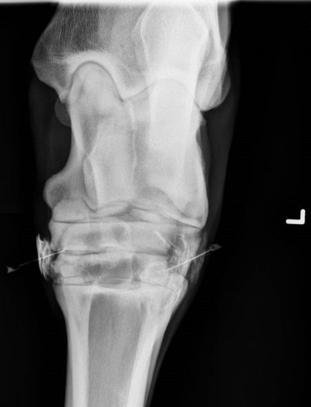
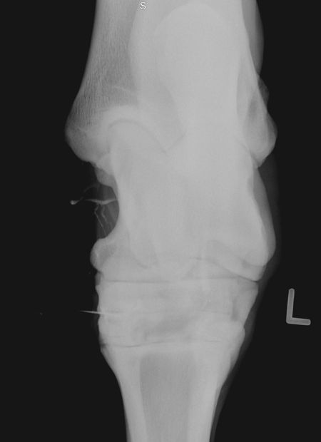
Tarsometatarsal joint. Palpate the head of the lateral splint bone, and insert a 1.5-in, 20-ga needle in a dorsomedial and distal orientation. Inject 3 ml of sterile radiopaque contrast material into the TMT joint first. Immediately after injection, and prior to obtaining radiographs, attach the PRN cap to the needle hub to prevent leakage of contrast and promote sterility. Obtain standard radiographic views and evaluate them for the presence of contrast medium in the PIT or TT joints (Figures 1A & 1B). If no proximal communication is seen, remove the PRN cap. Although not necessary, an empty syringe can be attached at this point to aspirate any excess contrast or synovial fluid present. Then, administer 3 ml of alcohol intra-articularly and remove the needle.

Figure 1A. A dorsoplantar radiographic projection after injection of contrast medium into the hocks. No radiographic communication between the DIT/TMT and PIT/TT is noted. As such, it was determined to be safe to inject the alcohol. (Figure courtesy of Dane M. Tatarniuk, DVM, MS, DACVS-LA)

Figure 1B. A dorsoplantar radiographic projection after injection of contrast medium into the hocks. Communication between the DIT and PIT/TT is present, as evident by contrast migrating proximally in the medial aspect of the TT joint. As such, it was determined not safe to inject the alcohol. (Figure courtesy of Chris D. Bell, DVM, DACVS, Elders Equine Clinic, Winnipeg, Manitoba, Canada)
Distal intertarsal joint. Contrast evaluation in the DIT should be repeated as described for the TMT joint (using 3 ml), regardless of the appearance of contrast arthrography after TMT injection (Figures 1A & 1B). The intersection between the fused first and second tarsal bone, the third tarsal bone and the proximally located central tarsal bone creates a palpable gap into which a 1-in, 22-ga needle can be inserted immediately distal to the central tarsal bone directly perpendicular to the skin in a medial to lateral orientation. If contrast arthrography continues to appear confined to only the DIT and/or TMT, then administer 3 ml of alcohol intra-articularly.
Proper technique requires the positive contrast radiographic assessment and subsequent alcohol injection in both the TMT and DIT independently. After the injection, horses receive a standard dose of phenylbutazone (2.2 mg/kg orally or intravenously). Some authors describe placing a hock bandage,4 but in my experience this is not necessary.
Cautions and complications
In one study, mild swelling and localized cellulitis at the site of injection was reported in 12.5 percent of cases.2 It has been suggested that difficulty with needle placement (more than two or three attempts to enter the joint) can create multiple leakage routes for alcohol or contrast medium to inflame the soft tissues.2 Depending on the volume administered extra-articularly, marked soft tissue fibrosis and undesirable periosteal reaction can develop, potentially worsening the lameness. Specific to the treatment of lower hocks, if contrast arthrography is omitted or incorrectly interpreted, injected alcohol may migrate through the PIT and into the TT joint. Since the TT joint is involved in a high degree of flexion and extension during gait movement, onset of iatrogenic arthritis secondary to alcohol diffusion will have crippling consequences.
Anticipated outcome
Retrospective studies evaluating alcohol-facilitated ankylosis have recently emerged. One study reported a significant improvement in lameness in all clinical horses evaluated after alcohol injection into the TMT and DIT joints (n = 21).4 Lameness improvement was also supported by objective force-plate analysis in half of these patients. Another distal hock joint retrospective documented an overall improvement in 63 percent of 35 treated limbs when the horses were re-evaluated subjectively six to nine months after injection.2 Alcohol-facilitated ankylosis of the proximal interphalangeal joint (pastern) has been successfully performed and is a viable option when other surgical interventions, such as pastern arthrodesis, are not an option.
Conclusion
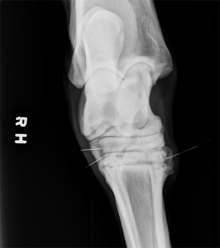
Alcohol-facilitated ankylosis of the DIT and TMT joints can be performed safely after contrast radiography using readily available equipment and standard hock injection techniques. Multiple injections may be necessary (Figure 2), and the technique is generally considered safe as long as the absence of communication with the PIT and TT joints is confirmed.

Figure 2: A dorsoplantar radiographic projection of a tarsus that was previously injected with alcohol intra-articularly eight months prior. Note the prominent callus present along the medial aspect of the TMT and DIT joints secondary to alcohol-augmented arthritic degeneration. In this case, ankylosis has progressed reasonably well along the medial aspect of the lower joints. However, a second round of alcohol is being administered to help promote complete fusion desired along the lateral aspect of the joint. (Figure courtesy of Dane M. Tatarniuk, DVM, MS, DACVS-LA)
Consult a board-certified equine surgeon prior to the procedure as more technical surgical interventions including thermal- (laser) or mechanical- (drilling with or without plating) facilitated ankylosis techniques may be more appropriate in select cases.
References
1. Shoemaker RW, Allen AL, Richardson CE, et al. Use of intra-articular administration of ethyl alcohol for arthrodesis of the tarsometatarsal joint in healthy horses. Am J Vet Res 2006;67(5):850-857.
2. Lamas LP, Edmonds J, Hodge W, et al. Use of ethanol in the treatment of distal tarsal joint osteoarthritis: 24 cases. Equine Vet J 2012;44(4):399-403.
3. Bell BT, Baker, GJ, Foreman JH, et. al. In vivo investigation of communication between the distal intertarsal and tarsometatarsal joints in horses and ponies. Vet Surg 1993;22(4):289-292.
4. Carmalt JL, Bell CD, Panizzi L, et. al. Alcohol-facilitated ankylosis of the distal intertarsal and tarsometatarsal joints in horses with osteoarthritis. J Am Vet Med Assoc 2012;240(2):199-204.

Dr. Dane Tatarniuk is an ACVS board-certified equine surgeon at Iowa State University in Ames, Iowa. He has a clinical and research interest in equine sports medicine, orthopedics, regenerative therapy and musculoskeletal traumatology. In his spare time, he enjoys camping and fishing, spending time with his wife, and helping his wife raise and show western pleasure Quarter Horses.

Surgery STAT is a collaborative column between the American College of Veterinary Surgeons (ACVS) and dvm360 magazine. To locate a diplomate, visit ACVS's online directory, which includes practice setting, species emphasis and research interests, at acvs.org.