Tick-borne disease in cats: Two to watch for
Vigilance is necessary for your feline as well as canine patients if you live in a tick-endemic areacytauxzoonosis and anaplasmosis can both affect cats.
Shutterstock.comThings are changing for feline vector-borne disase. If you thought you lived in a state where ticks weren't a problem, you might want to check again-they may be advancing into your area soon. Here are two that are much more problematic than they were in the recent past.
Cytauxzoonosis
Cytauxzoon felis is a protozoan blood parasite transmitted via the bite of an infected tick. Bobcats (Lynx rufus) are the reservoir host; infected bobcats are believed to develop short-lived illness followed by clinical recovery and a persistent carrier state. When a tick feeds on an infected bobcat, the tick acquires the pathogen. If that tick then feeds on a domestic cat, infection typically leads to severe illness (cytauxzoonosis) and often death. All Felidae are susceptible to infection; infection has not been reported in any non-felid animal.
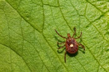
Amblyomma americanum, the lone star tick, is considered the predominant vector of C. felis based on experimental infection studies and epidemiologic data, though historical studies indicate that Dermacentor variabilis, the American dog tick, can also transmit C. felis to cats.
During feeding, the tick inoculates sporozoites that enter host mononuclear cells and multiply. Infected mononuclear cells distend with organisms known as schizonts. The multinucleated schizonts divide into merozoites. Within days, merozoites rupture from the mononuclear cells and are taken up by red blood cells (RBCs) where they appear as piroplasms. Widespread dissemination of schizonts results in parasitic thrombosis, circulatory impairment, tissue infection and a severe systemic inflammatory response, which can lead to multiorgan dysfunction and failure and death within three weeks of infection. Many cats die within 24 hours of presentation to a veterinary clinic for treatment.
In cats that survive initial infection, late-stage disease involves erythroparasitemia (piroplasm structures within RBCs) that can be readily observed in blood smears. Piroplasms persist for months, years or even for life in these cats, although schizonts can no longer be found. While clinically relevant hemolysis may occur in the acute phase of infection, chronic erythroparasitemia is relatively benign. The parasite life cycle is perpetuated when a tick takes a blood meal containing merozoite-infected RBCs.
It has now been documented that recovered domestic cats are competent to transmit the pathogen to feeding ticks, but C. felis cannot be transmitted through physical contact between infected cats. Although experimental inoculation of naive cats with schizonts results in cytauxzoonosis and death, inoculation of RBCs containing piroplasms does not lead to schizogeny or illness. Vertical transmission, common in other apicomplexan infections, has not been documented.
Since the first reported case of cytauxzoonosis was recognized in Missouri in 1976, its geographic range has progressively expanded. To date cytauxzoonosis has been confirmed in domestic cats in 17 states. In 2015, the first report surfaced of C. felis infection in Illinois. The pathogen has been documented in bobcats from Pennsylvania and North Dakota, states where ill domestic cats have not yet been reported (see map).
The increase in range is likely due to expansion in the range of the A. americanum population, hypothesized to be linked to an expanding white-tailed deer population. Veterinarians in areas where lone star ticks are found but cytauxzoonosis has not yet been recognized-including much of the northeastern and central United States-should be vigilant for the disease.
Veterinary practitioners in areas where cats are at risk should advise owners about disease prevention, including keeping cats indoors and using tick control products approved in cats. In most cats affected by cytauxzoonosis, initial disease signs include fever, anorexia and lethargy. Tachypnea and tachycardia are commonly observed. Within days, clinical signs can progress to severe weakness, icterus, respiratory distress and neurologic dullness.
Diagnosis can be made by identification of piroplasms within erythrocytes during microscopic examination of a peripheral blood smear. PCR assays have been developed to confirm the presence of C. felis and other Cytauxzoon species, but so far they are not useful as a quick diagnostic tool in practice. Still, it's recommended that samples from suspected cats be submitted to appropriate laboratories to further confirm the infection. Low levels of parasitemia can be detected only by PCR assay.
One clinical trial has demonstrated better survival rates (60% versus 26%) with the combination of atovaquone (15 mg/kg orally every eight hours) and azithromycin (10 mg/kg orally once daily) compared with imidocarb (3.5 mg/kg intramuscularly once) in 80 cats with acute disease.1 Supportive treatment was the same in all cats, consisting of fluid therapy and heparin. This study suggests that this antiprotozoal combination plus supportive treatment is the current approach of choice. In some cats nasoesophageal or esophagostomy tube may be needed to administer drugs and enteral feeding.
There is currently no vaccine against C. felis, so prevention is based on living indoors or use of effective tick treatment in cats with outdoor access. Efficacy of an acaricide collar (imidacloprid 10% plus flumethrin 4.5%) for prevention of C. felis transmission has been proven in a controlled prospective clinical trial.2
In the study two groups of cats (with collars and without) were exposed to A. americanum ticks infected with C. felis. No cats with a collar were infected, versus 90% of the cats without a collar.
Anaplasmosis
Anaplasma phagocytophilum is a rickettsial organism that causes granulocytic anaplasmosis in cats, dogs, horses, ruminants and humans. The worldwide distribution of A. phagocytophilum follows the geographic distribution of its primary vector, Ixodes species.
In North America, the organism is transmitted by Ixodes scapularis in the Northeast and Midwest and by Ixodes pacificus in the West. Infections are highest in the late spring and autumn when both nymph and adult ticks are most mobile. Transmission to mammals occurs within 24 to 48 hours of tick attachment.
Once transmitted, A. phagocytophilum infects circulating neutrophils, forming intracellular inclusions (morulae) that can be observed via light microscopy on a Romanowsky or Wright-Giemsa-stained blood smear. In North America, morulae have been identified in the neutrophils of naturally exposed dogs, horses and humans and experimentally infected cats. In Europe, morulae of A. phagocytophilum have been described in ruminants, cats, horses and dogs.
In a recent retrospective study researchers identified 16 cases of anaplasmosis in cats.3 The objectives of this study were to describe the clinical and historical findings in cats that were positive by PCR for A. phagocytophilum DNA in their blood and to describe treatment and response. Investigators were also able to characterize intracellular morulae identified in neutrophils on microscopic examination of peripheral blood smears.
Of the 16 cats, all were lethargic and 15 had a fever. Fourteen were anorexic. Other signs were variable and may not have been specific for Anaplasma infection. All had access to the outdoors and lived in Ixodes-endemic regions. Treatment information was available in 15 cats. Another antibiotic had been prescribed prior to PCR amplification of A. phagocytophilum in eight cats. Once PCR assay results confirming the presence of A. phagocytophilum DNA were available, all 15 cats were administered doxycycline orally.
Clinical abnormalities resolved after initiation of doxycycline therapy in the 14 cats whose response to treatment was known. The duration of doxycycline administration varied from 21 to 45 days, with most cats treated for 21 days. Previous reports in cats have recommended treatment with 5 mg/kg doxycycline orally every 12 hours for 28 days, but the ideal duration of treatment with doxycycline in cats is unknown and warrants additional study.
A phagocytophilum infection should be included on a differential diagnoses list for any cat that lives in an Ixodes species–endemic area with potential tick exposure and presents with vague clinical signs (either acute or intermittent) of lethargy, fever and anorexia. A phagocytophilum infection can be identified by documenting DNA in peripheral blood by PCR prior to antibiotic administration or by identifying morulae within neutrophils on a direct blood smear examination. Exposure can be documented by demonstrating the presence of antibodies with ELISA and indirect fluorescent antibody serology; a fourfold change in convalescent titers after 14 days indicates an active infection. Year-round tick prevention should be included to preventive care for cats living in Ixodes-endemic areas.
References
1. Cohn LA, Birkenheuer AJ, Brunker JD, . Efficacy of atovaquone and azithromycin or imidocarb dipropionate in cats with acute cytauxzoonosis. J Vet Intern Med 2010; 25:55-60.
2. Reichard MV, Thomas JE, Arther RG, . Efficacy of an imidacloprid 10 %/flumethrin 4.5% collar (Seresto, Bayer) for preventing the transmission of Cytauxzoon felis to domestic cats by Amblyomma americanum. Parasitol Res 2013; 112 Suppl 1:11-20.
3. Savidge C, Ewing P, Andrews J, et al. Anaplasma phagocytophilum infection of domestic cats: 16 cases from the northeastern USA. J Feline Med Surg 2016 Feb;18(2):85-91.
Further Reading
1. Lloret A, Addie DD, Boucraut-Baralon C, et al. Cytauxzoonosis In cats: ABCD guidelines on prevention and management. J Feline Med Surg 2015 Jul;17(7):637-41.
2. Sherrill MK1, Cohn LA. Cytauxzoonosis: Diagnosis and treatment of an emerging disease. J Feline Med Surg 2015 Nov;17(11):940-8.
Dr. Elizabeth Colleran is owner of Chico Hospital for Cats in Chico, California, and a frequent speaker at the CVC conferences.
Newsletter
From exam room tips to practice management insights, get trusted veterinary news delivered straight to your inbox—subscribe to dvm360.


