Why Aren't my Patients Responding to Their Allergy Shots?
I often get referrals of patients on immunotherapy that "aren't doing well" or hear from the owner that "those shots never helped so we stopped them."
I often get referrals of patients on immunotherapy that "aren't doing well" or hear from the owner that "those shots never helped so we stopped them."

A young, atopic Irish Wolfhound before (left) immunotherapy and responding (right) to immunotherapy.
The referring veterinarian has performed in vitro testing or skin testing and started the patient on immunotherapy based on the test results. What went wrong? Why didn't the injections help the patient? What can you do to help make those injections be more successful?
1. Is the diagnosis of atopy correct?
It sounds elementary, but there are other diseases that mimic atopy because they involve the same areas of the body, start between the ages of 6 months to 3 years, and are steroid responsive. Before the diagnosis of atopy can be made, rule-outs of flea allergy or other ectoparasites such as Cheyletiella or scabies, food allergy, bacterial pyoderma and Malassezia dermatitis must be eliminated.
In essence, atopy is a diagnosis of exclusion. Most atopics begin with pruritus or recurrent pyoderma between the ages mentioned above (6 months to 3 years), are of a certain breed due to atopy being hereditary (most commonly Retrievers, Shepherds, Terriers, Dalmatians, herding breeds, Poodles), are responsive to steroids at antipruritic doses (0.25 mg/lb/day), and most often involve face and feet (foot licking, recurrent ear infections, face rubbing and occasionally rectal pruritus). It is important to rule out ectoparasites such as fleas and other contagious mites as the first step in working up a patient suspected to be atopic.

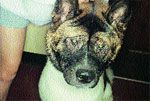
Periocillar alopecia due to face rubbing in an atopic Akita.
2. Are bacterial pyoderma or Malassezia dermatitis complicating the allergy?
This often occurs in a patient that had previously been under good control with immunotherapy for their atopy but perhaps "breaks out" during their "worst" affected season. One solution to prevent this from occurring next time is to increase the particular allergen(s) that the patient is reacting to.
For example in Cleveland, ragweed season begins mid-August and runs through mid-October. If the patient flares with a Malassezia otitis/dermatitis or a bacterial pyoderma during this time and is ragweed allergic, we need to increase the amount of ragweed in the allergy solution to help better "protect" him. At the same time, the pyoderma must be addressed using antibacterial bathing and an antibiotic with activity against Staph intermedius such as cephalexin, lincomycin, enrofloxacin, amoxicillin/clavulanic acid or combination sulfas. Yeast problems should be treated with an antiyeast shampoo and oral ketoconazole if necessary. To determine whether yeast or bacteria are present, skin cytology and ear smears should be performed and the patient treated appropriately.
3. The patient is more pruritic with each subsequent allergy injection.
Remember you are giving the patient what it is allergic to so an injection that is "too strong" will actually cause more pruritus. If the patient was administered 0.5cc of a 2000pnu solution and the owner feels he was itchier the entire week with pruritus improving the farther away they were from the injection, the amount administered must be reduced. In this situation, we would reduce the dose to 0.4cc and note the response. Unfortunately immunotherapy is not a matter of administering the weekly injections per the instruction sheet. Attention must be paid to the patient's degree of pruritus following that injection. If pruritus increases after the injection, the dose should be reduced, if no reduction in pruritus, keep going up on the shots. Occasionally the owner feels the "shots aren't doing anything". In that case we will actually skip a week on the injections to see if he/she is less itchy without the shot (means the dose was probably too high) or more pruritic after missing a dose (means the shots are doing something, but we need to go higher). Remember the goal is not to get to the final dose of the strongest allergy vial but to whichever dose of whichever vial keeps that patient under control.

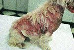
Atopic West Highland White Terrier with a secondary malassezia dermatitis. Note: Erythema, alopeica and seborrhea.
In some cases, the very first injection makes the patient extremely pruritic. In that case, we will dilute the solution down ten-fold and start at 0.1 cc/wk.
Finally, there are occasional patients that become pruritic after an injection just from the very act of receiving an injection! We usually premedicate those patients with an antihistamine such as hydroxyzine l mg/lb, 30-60 minutes prior to the injection.
4. Be sure the allergy test results match the time of the year the patient is affected.
Once you have determined via patient history and clinical signs that the patient is atopic, skin or in vitro testing is performed to determine which allergens go into the allergy solution. When the results are obtained, the positive allergens must correlate with the time of the year the patient is affected.
For example, in the Midwest where we have specific pollen seasons i.e. spring-trees, summer-grasses, fall-weeds, it can sometimes be predictable as to which group the patient will be allergic. However in warmer climates where pollination occurs almost year round, it can be more difficult to "match" the allergens to the patient.

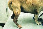
A dog with signs of chronic atopy - lichenification, hyperpigmentation and alopecia.
Nonseasonal atopics may be dust mite sensitive. However keep in mind that some Malassezia dermatitis patients or patients with ectoparasite infestations such as Cheyletiella or scabies will skin test positive to house dust mite antigen. Rather than just checking off all of the positives on the in vitro test for the allergy company to place in the solution, sit down and review the patient's chart to see what time of year they are affected and if, in fact, that is a particular pollen season in your area.
5. The patient is allergic to "everything".
After reading No 4. above, if it really is true that he/she is allergic to many trees, weeds, grasses, etc. keep in mind it is best not to include >12 allergens in one vial. Some patients need two separate vials because they are allergic to so many antigens.
Also if your patient is on a vial that includes many allergens and they are up to the maximum dose and doing better but not as well as you'd like, you may need to have the vial made stronger.
For example if the patient is on l cc/wk of a 20,000-pnu vial and improves after his weekly injection but it doesn't seem to "hold" him for but a few days, you may want to request a 30,000-pnu solution.

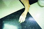
Acral lick granuloma in an atopic Labrador Retriever and erythemic caudal aspects of the feet due to licking.
Molds should not be mixed in the same vial as grasses or weeds. Molds produce a protease enzyme that can inactivate these pollens. If a patient is mold allergic as well as pollen allergic, we will usually make up a separate mold only allergy solution as well as a separate pollen solution. Interestingly, the group of allergens that most often produces false positive reactions on in vitro testing is fungi or molds. The question then becomes is the patient truly mold allergic or are these false positive values?
6. Skin testing or in vitro testing do not support a diagnosis of atopy.
False negative skin or blood tests for atopy can occur if the patient is tested away from their affected season.
For example in the Midwest if we skin test a patient in the middle of winter and he is tree or grass allergic, the test may not pick up on this and we will need to retest at a different time of the year. Fall seems the best season to skin test since the patient just experienced tree (spring), grass (summer), and weed (fall) pollens.
Was the patient off steroids long enough to register positive on testing? Some patients are extremely steroid sensitive and the use of topical steroids alone may inhibit positives on both skin and in vitro testing. Because of this individual and unpredictable steroid sensitivity, each patient must be evaluated to see how long they need to be off steroids before testing. Factors such as how long the patient has been on steroids, at what intervals, and whether short-acting or long-acting steroids were used all need to be taken into consideration. Generally six to eight weeks off steroids is usually adequate in most patients. Antihistamines usually do not interfere with in vitro testing but must be discontinued seven to 14 days prior to skin testing.
Unusual but true in some atopic patients is that some skin test positive and in vitro test negative while others do the opposite. We generally skin test all of our atopic patients but occasionally we will get a negative skin test yet the patient registers positive on in vitro testing.
7. Close communication with the client is essential.
This is true especially when beginning the allergy injections. Once the patient's maintenance dose is achieved then the frequency of the injections can be altered.
However since immunotherapy can take three to six months, and sometimes up to a year, the start of allergy injections can be trying for the owner (and the doctor!) because anxious owners may want to give up because they are not seeing results. Communication via handouts and phone conversations can be helpful in explaining and giving encouragement. Be sure to do one thing and one thing well... listen to the owner! They live with the patient and know how pruritic he/she is before and after an injection. It is very important that they be observant. Our next decision is based on their feedback to us and ultimately determines how well the patient fares.
Newsletter
From exam room tips to practice management insights, get trusted veterinary news delivered straight to your inbox—subscribe to dvm360.
Zenrelia™ (ilunocitinib tablets): An Innovative, Once Daily Canine Treatment That Targets Itch
June 25th 2025Finding an effective, safe medication to control itch is essential to maximize patient comfort. Because Zenrelia does not require a loading dose and is administered once daily, Zenrelia may help improve owner compliance and can be cost effective* for pet owners
Read More