3 unusual (but not uncommon) skin diseases you should know
These three skin diseases arent zebrastheyre more like donkeys. Learn to recognize their hoofbeats so you dont miss the diagnosis.
You've probably heard the saying, “When you hear hoofbeats, think horses not zebras.” But what if you turn and find a donkey?
I like to think of pemphigus foliaceus, calcinosis cutis and hepatocutaneous syndrome as the “donkeys” of skin diseases. Most veterinarians don't see them every day, but they are not uncommon. You will likely see patients with these diseases, and if you confuse them with horses or zebras, you might feel like an ass (pun intended).
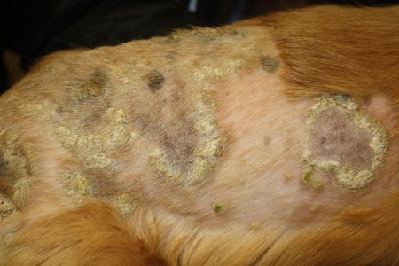
Pemphigus foliaceus (All images courtesy of Dr. Darin Dell)Skin disease donkey #1: Pemphigus foliaceus
Pemphigus foliaceus is the most common autoimmune skin disease in dogs. Out of the three skin disease donkeys, it is also the most likely to be seen by a general practitioner.
Pemphigus foliaceus affects the two uppermost layers of the epidermis. It is typically idiopathic but can develop secondary to drug exposure. Pemphigus foliaceus triggered by metaflumizone (ProMeris) has been well documented, and cimetidine (Tagamet-Prestige Brands) and various antibiotics have also been implicated.
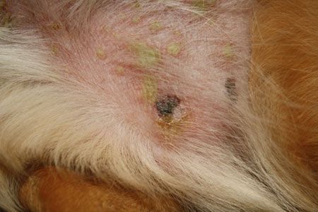
Pemphis foliaceusLesions
Pemphigus foliaceus is characterized by pustules and honey-colored crusts. These pustules are large and fragile and can develop quickly-sometimes in less than an hour. They are commonly found on the face and trunk, although they can be anywhere. Discreet pustules may be seen on the paw pads, but thickened, dry and crusted pads are more common.
Diagnosis
To definitively diagnose pemphigus foliaceus, submit a biopsy sample. If possible, intact pustules are preferred, and be sure to include crust debris.
Treatment
Corticosteroid therapy will successfully treat pemphigus foliaceus in most patients, but it is a lifelong therapy. While dogs will initially require daily dosages, over the course of three or four months, most can be tapered down to dosages every other day.
Most patients also need to receive a corticosteroid-sparing agent (e.g. cyclosporine, azathioprine) to reduce the side effects of corticosteroid therapy. Since it takes four to eight weeks for these drugs to be effective, I typically give patients a corticosteroid and corticosteroid-sparing drug together from the beginning of treatment.
Secondary bacterial infection is common in these patients. Treat with oral antibiotics as needed.
Pro tips
1) Even the slightest disturbance to the skin can damage these pustules, so don't scrub the patient's skin or shave its fur before collecting samples.
2) These patients need to be re-examined frequently (every two to three weeks) when you start therapy so you can evaluate the crusts and monitor for medication side effects.
3) Clients won't be able to distinguish a crust caused by infection from a crust caused by pemphigus, so check cytology at each visit.
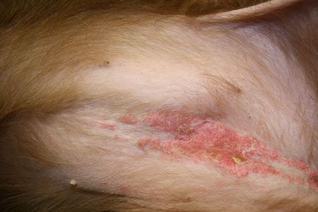
Calcinosis cutisSkin disease donkey # 2: Calcinosis cutis
Calcinosis cutis is characterized by calcium salt deposits in dermal and epidermal tissues and is the second most common of the three skin disease donkeys. Because it is often linked to corticosteroid use, whether or not you see it frequently in your practice depends on your area's veterinary culture.
Causes
Common cause: hyperglucocorticoidism, secondary to corticosteroid administration or hyperadrenocorticism
Less common cause: secondary to percutaneous calcium absorption (e.g. contact with an ice melt product containing calcium chloride such as MELT or Road Runner Ice Melt, or calcium fertilizers such as CalMax or Foli-Cal)
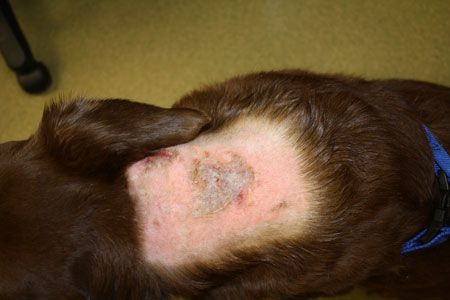
Calcinosis cutisLesions
Early lesions are chalky white to pink with indistinct margins. Advanced lesions are white and firm and are usually surrounded by redness and swelling. For some pets, these are very painful. Lesions typically develop on the dorsal neck and then spread caudally. Any dog can develop these lesions, but they are most commonly seen in English bulldogs and dogs receiving corticosteroid injections.
Diagnosis
Calcinosis cutis can be seen with radiography. But to definitively diagnose this disease, collect biopsy samples from areas not affected by self-trauma and ulceration. Biopsy results reveal diffuse or multifocal calcification of dermal collagen.
Treatment
Stop corticosteroids and treat secondary infections and any systemic disease. Calcium deposits will dissolve in two to 12 months.
Pro tips
Patients with calcinosis cutis do not have elevated serum calcium concentrations.
Patients with a history of calcinosis cutis should never receive corticosteroid therapy again.
Dimethyl sulfoxide (DMSO) gel can slightly reduce the time it takes for the calcium deposits to dissolve. However, I don't recommend it because most owners can't tolerate the smell.
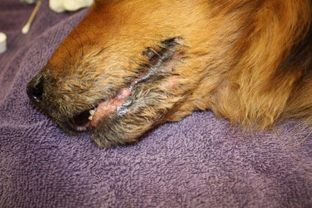
Hepatocutaneous syndromeSkin Disease Donkey # 3: Hepatocutaneous syndrome
Hepatocutaneous syndrome is the least common of the three, and that is fortunate because it carries a grave prognosis. Typically seen in senior dogs, the disease involves the death of keratinocytes in the epidermis due to amino acid starvation. Most affected dogs have a distinctive chronic hepatopathy.
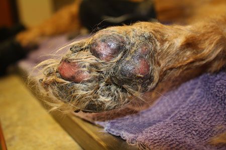
Hepatocutaneous syndromeLesions
Skin lesions are typically the first sign, rather than signs of liver disease. Crusts and erosions occur most commonly on paw pads, elbows, hocks and muzzles. Many patients are reluctant to walk because of damage to the paw pads.
Diagnosis
A complete blood count, serum chemistry profile and urinalysis are recommended. Nonregenerative anemia and hyperglycemia are common. The serum chemistry evaluation may not reveal any abnormalities.
Abdominal ultrasonographic examination results will likely show a liver with a honeycomb pattern, but inexperienced ultrasonographers may misinterpret the liver changes. Biopsying the skin is the ideal method for diagnosing this condition. However, if your patient is a high anesthetic risk, ultrasound alone may be sufficient. In an ideal situation, I recommend both. Collect biopsy samples of affected skin with attached crust. Do not biopsy ulcerated or eroded tissue.
Treatment
Hepatocutaneous syndrome is a marker of severe internal disease. Consider referring these patients to a dermatologist and to an internist.
Therapy with a crystalline amino acid solution (Aminosyn-Pfizer) can replenish the amino acids the patient is missing. These injections are typically given once or twice a week initially. Once the patient has shown a good response, the injections are administered every four to eight weeks long term. Aminosyn must be given intravenously over an extended period so hospitalization is required. The therapy is expensive, but it may yield a clinical response for up to 22 months.
Because of the expense of Aminosyn and the poor prognosis, most owners opt for supportive therapy, such as increased intake of protein and fatty acids. Corticosteroid therapy can temporarily improve clinical signs as well, but patients eventually become resistant. The client should be warned that corticosteroid administration in this situation greatly increases the risk of diabetes mellitus. The typical life expectancy with supportive therapy alone is two to five months.
Secondary infections are a constant concern for these patients because their skin does not heal well. Many of these patients do not eat well either, so I recommend treating with antiseptic sprays and wipes rather than oral antibiotics.
Pro tip
These lesions show up on the places of the body with the most wear (e.g. paw pads, elbows, muzzle) because that is where the skin needs to be renewed most frequently and where it requires the most nutrition. Thus, lesion location can help differentiate this disease.
Dr. Darin Dell spent six years in general practice and two years in emergency medicine before becoming a Diplomate of the American College of Veterinary Dermatology in 2012. He is currently on staff at Animal Dermatology Clinic in Indianapolis. Dr. Dell's hobbies include woodworking and mountain biking.