The ABCs of veterinary dentistry: 'R' is for retained, primary, deciduous teeth
Attention to persistent primary teeth is essential to the dental health of our patients, especially smaller breeds such as Maltese, Yorkshire terriers, Pomeranians and miniature Schnauzers.
In cats and dogs, primary (baby) tooth roots are normally resorbed from pressure as the permanent (secondary, adult) teeth erupt pushing them out of the alveolus, starting at 14 weeks of age. The mechanism that causes resorption of primary roots isn't fully understood, nor is the cause of resorption failure. Persistent primary teeth, fail to exfoliate because the permanent tooth buds are malpositioned rostrally (maxillary canines) or lingually (mandibular canines) removing the direct force to push them out of mouth. Permanent canines normally erupt by the time most dogs and cats are 6 months old.
Defining the problem
Let's take a look at the terminology of this dental issue.
Primary-The first teeth, which are normally shed and replaced by permanent teeth.
Retained-Primary teeth that continue to be present in cases where secondary teeth are not present.
Persistent-Primary teeth that are still present despite the eruption of permanent teeth.
Deciduous-A dental term applying to the primary teeth that is borrowed from trees and shrubs that seasonally shed leaves as a tree matures.
Secondary-Adult teeth.
The terms “retained deciduous” and “retained primary” should be reserved for rare cases when only the primary tooth clinically and radiographically exists without an accompanying secondary (adult) tooth. (Figures 1A-1C).
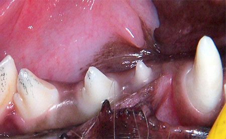
Figure 1A. A retained right mandibular primary second premolar. (All photos courtesy of Dr. Bellows.)
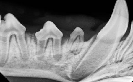
Figure 1B. A radiograph confirming the absence of secondary first and second premolars.
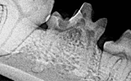
Figure 1C. A retained deciduous fourth premolar.
Persistent primary teeth are diagnosed when the primary and secondary teeth are present in the same alveolus. This results when the normal resorption of primary teeth fails to occur due to malposition of the secondary tooth, causing the secondary teeth to erupt next the primary teeth. A retained deciduous tooth occurs where there is a primary (deciduous) tooth without an accompanying secondary (adult) tooth visible either clinically or radiographically (Figures 2A-2E).
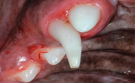
Figure 2A. A persistent right maxillary canine. Note the swelling around the primary and secondary canine.
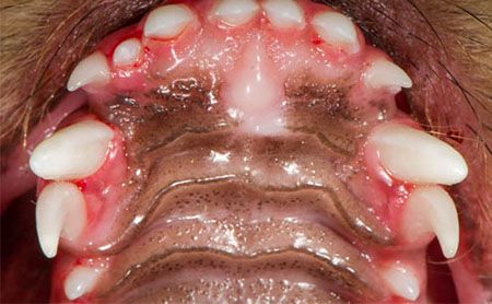
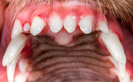
Figures 2B and 2C. Persistent primary maxillary canines and the right third incisor.
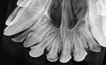
Figure 2D. Radiographic confirmation of the persistent right maxillary third incisor.
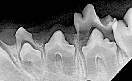
Figure 2E. Persistent primary left mandibular second and fourth premolars.
Persistent primary teeth may overcrowd the dental arch, moving the secondary teeth to abnormal locations, causing oral discomfort. Double sets of roots may also prevent the normal development of the alveolus and periodontal support around each permanent tooth, resulting in early tooth loss. Malpositioned, primary mandibular canine teeth result in mesioversion (lingual displacement) of the permanent mandibular canine teeth causing traumatic occlusion of the hard palate (Figures 3A and 3B).
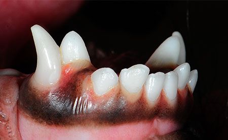
Figure 3A. A persistent primary right mandibular canine producing mesioversion of the secondary mandibular canine.
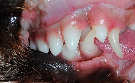
Figure 3B. Mesioversion of the left mandibular canine.
When a delayed approach is taken to determine whether the persistent primary tooth will exfoliate, the secondary adult tooth often becomes permanently malpositioned, requiring orthodontic movement, crown reduction or extraction. It is for this reason that the “wait and see” approach isn't recommended.
Just say 'no' to a trim
Some breeders trim the primary canine crowns in hopes that they'll shed early and possibly prevent orthodontic problems. Trimming, also known as deciduous tooth crown reduction, isn't recommended because it results in pulp exposure, causing the animal pain and risking the development of the surrounding permanent teeth.
Treatment
Now that we've defined the problem, let's fix it! A persistent primary tooth should be extracted as soon as the permanent tooth is observed to erupt in the same alveolus. The goal is to remove the entire primary tooth without fracture of the root. Examination of intraoral radiographs before extraction is important to get an appreciation of the subgingival anatomy of the tooth to be extracted.
Here are the extraction steps after examining intraoral radiographs:
1. Make a diagonal incision over the caudal primary canine root (Figures 4A and 5A).
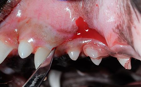
Figure 4A. Left maxillary persistent primary canine tooth. Gingical incision used to expose the persistent primary tooth.
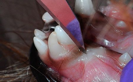
Figure 5A. Left mandibular persistent primary tooth extraction indicated.
2. Use a No. 2 molt periosteal elevator to expose the primary canine root (Figure 4B).
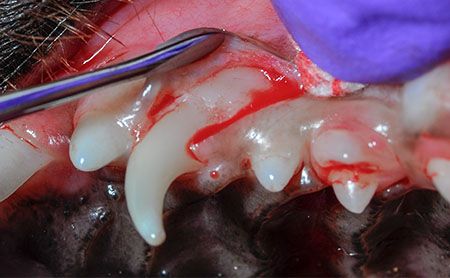
Figure 4B. Flap exposure of the primary canine tooth.
3. Insert and gently torque a wing-tipped elevator to create mobility of the tooth before delivery (Figures 4C and 5B).
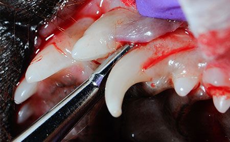
Figure 4C. Wing-tipped elevator used to loosen the primary canine tooth.
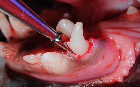
Figure 5B. Wing-tipped elevator used during extraction.
4. Use extraction forceps or a rongeur to deliver the tooth from the alveolus (Figures 4D and 5C).
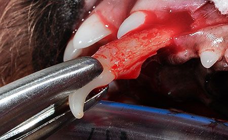
Figure 4D. Primary canine tooth delivered from the oral cavity.
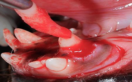
Figure 5C. Persistent primary tooth delivered from the oral cavity.
5. Suture the incision with 4-0 absorbable suture on a P-3 reverse cutting needle.
Extraction must be done carefully to avoid accidental damage to the unerupted, permanent canine tooth that lies lingual to the mandibular teeth and rostral to the maxillary deciduous canines. Avoid placing the elevator along the lingual surface of the mandibular deciduous teeth. Instead, only elevate along the mesial surface (front), labial surface (toward the lip), distal surface (caudally) of the mandibular deciduous teeth and buccal distally around the maxillary deciduous canines. If extraction is performed early, the abnormally positioned permanent tooth frequently moves into the normal position.
Dr. Jan Bellows owns All Pets Dental in Weston, Florida. He is a diplomate of the American Veterinary Dental College and the American Board of Veterinary Practitioners. He can be reached at dentalvet@aol.com.