Acute and chronic renal disease (specifically in lizard species) (Proceedings)
Kidney related diseases are a major cause of illness and death in captive lizards.
Kidney related diseases are a major cause of illness and death in captive lizards. Improper captive husbandry and diet are the most common predisposing causes of chronic renal failure - which is typically seen in adult lizards. Whereas acute onset of renal disease is often due to infectious or toxic causes (Including medications) and appears to effect any age animal and is typically more sporadic in occurrence. Because historical and clinical presentation can vary, the veterinarian must often rely on hematology and biochemistry, urine analysis, radiography, and biopsy to arrive at a definitive diagnosis.
The goal of this presentation is to provide the practitioner with an overview of renal disease and discuss options to arrive at a definitive diagnosis and determine an appropriate treatment plan.
To properly understand and appreciate the problems of renal disease in lizard species - a few basic anatomic and physiologic peculiarities must be understood. All lizards are essentially uricotelic, in that their primary excretory product of protein metabolism are salts of uric acid (urates) which are produced by the liver. Uric acid is largely insoluble which serves to reduce insensible water losses associated with excretion but does predispose dehydrated animals to gout if plasma uric acid levels rise above 63.73 mg/dl (1487 mol/L)1. Uric acid is actively secreted via the proximal renal tubules. The squamate kidney is metanephric in structure having relatively few nephrons, no loop of Henle nor a renal pelvis. Therefore, most reptiles are virtually unable to concentrate urine of a greater osmolarity than that of plasma. In addition, sexually mature male lizards may possess a sexual segment proliferation of the distal tubule. This is a pale pink glandular tissue designed for the production of seminal fluid. The gross appearance of the kidneys is usually dark brown/burgundy in color, they are paired, short and broad with few lobes. They are typically located in the caudodorsal coelomic cavity or pelvis. In laterally flattened lizards (i.e., chameleons) the kidneys are located craniodorsal to the pelvis.
In lizards, the urine produced by the kidneys flows down a ureter-like mesonephric duct to the urodeum of the cloaca, where it then passes into the bladder (if present) or cranially into the distal colon for storage prior to evacuation. Variable changes in urine concentration and electrolyte composition can occur within the bladder or distal colon. This means that bladder urine is not sterile and may not be a true osmotic/electrolyte representation of renal urine. A renal portal system is present but its functional significance is questionable1.
The renal portal system of reptiles allows for venous circulation from the hindlimbs and tail to course directly to the kidneys. Because of this, it has been recommended that medications should not be injected into the hind legs or tail as they will be conveyed to the kidneys, increasing the possibility of nephrotoxicity for aminoglycosides, and in the case of other drugs, reducing the likelihood of achieving the desired therapeutic effect. However, it is believed that a valve does exist, similar to that described in birds, shunting the blood, allowing it to pass either through the kidneys, or directly from the renal portal system to the postcava, bypassing the renal parenchyma.
Using fluoroscopy, one study demonstrated a structure histologically compatible with a valve was observed within the lumen of the abdominal vein. When the valve is closed blood would be forced into the iliac vein and enter the kidney and when it is open blood would enter the abdominal vein and bypass the kidney1. Due to the fact that only limited pharmacokinetic studies on the effect of the renal portal system on serum drug concentrations have been done, and considering the potential for nephrotoxicity with certain medications - it is still recommended to give all injectible medications in the first 1/2 to 1/3 of the body in reptiles.
History and Physical Examination
The history of illness in acute renal disease is usually relatively rapid onset of depression, lethargy, anoria, and weakness, often with a complete cessation of urate output. A complete history may determine the prior use of nephrotoxic drugs (i.e. aminoglycosides) or exposure to toxins or poisons. Frequently these animals were well maintained with a good level of nutrition and a reasonable level of husbandry. Therefore, on physical examination, most cases of acute renal disease will present in good weight and reasonable body condition. Dehydration may be evident due to reduced skin elasticity or decreased oral mucosal hydration, and pharyngeal or palpebral edema may be noted. The kidneys may or may not be palpable, with or without coelomic/abdominal tenderness or fluid accumulation.
In chronic renal disease, there will often be husbandry (low humidity, mild long term water deprivation/chronic low grade dehydration) or nutritional factors (high protein diets, excess vitamin D3 supplementation) that may indicate the potential for renal compromise. These animals tend to present with a history of reduced appetite, poor weight gain or weight loss, and occasionally the owners may report increased drinking in the animal. On physical exam the animals are usually of poor body condition, dehydrated and the kidneys may or may not be palpated either externally, or internally in larger animals, with or without abdominal tenderness. Unfortunately, most owners do not recognize the initial signs associated with renal disease, and so chronic cases will often be presented to the veterinarian as emergencies just like the acute renal failure case.
Though a complete physical exam is essential, it is often necessary to incorporate laboratory techniques and ancillary diagnostics to arrive at a definitive diagnosis.
Laboratory Techniques and Data
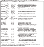
A basic data base including a complete blood count and serum biochemistry is essential in any reptile presenting with signs of anorexia, lethargy and depression - and will direct the veterinarian towards further laboratory tests and ancillary diagnostic procedures (Table 1).

Table 1. Blood Parameters in Green Iguanas, Iguana iguana, Used in Assessment of Renal Disease
1. The calcium and phosphorus ratio is often used as a reliable indicator of renal disease, and has been found to increase before any other biochemical parameter. 2. Calcium and phosphate ions, when present at appropriate concentrations, form insoluble salts in aqueous solutions. The activities of these ions required for precipitation to occur are determined by the solubility product constant (SPC) of the insoluble mineral. 3. The solubility product constant (solubility index) is calculated as the product of Ca (mg/dl)/{mmol/L} x PO4 (mg/dl)/{mmol/L}, and is normally less than (55)/{9}. If the solubility index rises above (70)/{12} then healthy tissue will start to mineralize, while between (55 - 70)/{9 - 12} mineralization of diseased tissue (i.e. kidneys) will likely occur.
Though often not sterile, urine samples, either freshly voided or obtained via cystocentesis, should be examined. Microscopic examination may reveal blood, a high inflammatory presence or renal casts indicating active infection and acute disease. Again, though normal bladder urine may not be sterile - if a culture and sensitivity reveals a profuse growth of a single organism, then that bacteria may be significant and appropriate antibacterial treatment is indicated.
Further Diagnostics: Imaging and Endoscopy
Dorsoventral radiographs are useful in most lizards for assessing kidney size, especially if the kidneys are enlarged. Radiography can also be used to demonstrate the presence of renal calculi and/or soft tissue mineralization. laterally compressed lizards (i.e. chameleons) a horizontal beam x-ray is more useful. Other important changes to look for are bladder stones, changes in bone density and constipation due to renal enlargement impinging on the pelvic canal. Intravenous urography can be very useful when attempting to identify renal changes (abscesses, neoplasia, calculi), renal and ureteral damage and ureteral obstructions. Via cephalic or jugular catheter 500 mg/kg of suitable water soluble iodine compound is injected. Some success has also been reported using intraosseous catheters in the proximal tibia. Radiographs are then taken at 0, 5, 15, 30 and 60 minutes post injection as needed to monitor the clearing of dye into the urinary system.
Ultrasonography from the ventral mid line and just caudal to the vent can also be employed to assess gross pathological renal changes. As mentioned earlier, MRI, which is superior to CT scanning for evaluating soft tissues, can be used - however, because of limited availability and cost - such imaging techniques are not often employed by general practitioners.
Ultimately, the veterinarian is expected to make a definitive diagnosis, determine if it is acute or chronic renal disease, provide specific therapy and general supportive care, and to provide the owner with an accurate prognosis. To this end, it has been found that the renal biopsy represents the most important diagnostic and prognostic tool when evaluating renal disease.
There are several techniques for collecting renal biopsy samples. Four techniques are: major celiotomy approach, cranial tail cut-down approach, transcutaneous needle biopsy and endoscopic biopsy. In the vast majority of cases the biopsy samples taken will provide the veterinarian with a definitive diagnosis and help to then give the owner a prognosis.
Treatment for Acute and Chronic Renal Disease
In acute renal disease/failure - the goal is to keep the animal alive until sufficient healing and recovery have taken place. Though the initial prognosis may be guarded - if imbalances are corrected, the chance for complete recovery does exist. An accurate weight of the animal is taken as is the packed cell volume (PCV) and total plasma protein - in an effort to accurately assess hydration status. Rehydration using o.18 % saline + 4 % glucose is recommended at 20 - 40 ml/kg/day i.v. or i.o. Lactated ringers solution (Hartmann's solution) may be less appropriate in cases of hyperkalemia (> 8 mmol/L)2. There is concern that significant acidosis may exist in acute renal failure - and this should potentially be addressed. In the past, acidosis has been ignored in the hopes that the acid-base disturbance would be resolved as renal perfusion and function was restored and the kidneys then corrected the acidosis.
The hydration status of the animal should be monitored using serial weight and PCV measurements. When correct hydration has been achieved, it is vital that over hydration be avoided and therefore a reduction in maintenance fluids to 2 - 10 ml/kg/day is required - but hydration status must continue to be monitored. If over hydration does occur, as evidenced by pharyngeal and pulmonary edema, then the use of diuretics is advisable (i.e. furosemide, thiazides)2.
In cases where uric acid levels are significantly elevated ( > 15 mg/dl / 750 mmol/L) the use of allopurinol (20 mg/kg p.o. q 24 hr) may reduce hepatic uric acid production, while the administration of anabolic steroids may reduce protein catabolism. In specific cases of pre-renal acute renal failure, rehydration, restoration of circulatory volume and supportive therapy may be all that is needed. In cases of post-renal obstruction, renal calculi and ureteral obstructions - these conditions will often have to be treated surgically before proper urine flow can be re-established.
In cases of toxin induced nephropathy - identification and removal of the toxin from the environment and gastric lavage may be useful. Where aminoglycoside toxicity is suspected - all medicating should stop and proper osmotic diuresis initiated to rehydrate and maintain appropriate renal perfusion. Acute hypercalcemia (i.e. from acute vitamin D3 overdose, not in breeding females) can result in acute ischemic tubular necrosis through the development of nephrocalcinosis, and in such cases it has been reported that prednisolone, calcitonin and diuresis should be considered2. Chronic renal damage can also lead to calcium salt deposition in soft tissues including the kidney due to an elevation in the solubility index. And, acute renal disease due to infectious agents should be empirically treated with broad spectrum antibiotics until culture and sensitivity results are obtained. It is very important to use medications with a large safety margin as drug metabolism and excretion may be significantly affected.
Once hydration and any underlying causes have been addressed - if the lizard still remains oliguric - then i.v. or i.o. administration of 20 % dextrose may be used in an attempt to induce diuresis. Initally the dextrose is given at o.4 - 1.0 ml/kg/hr for 30 - 60 minutes, then the rate is reduced to o.2 - o.5 ml/kg/hour. If the lizards continues to remain oliguric, then diuretics and coelomic dialysis may be attempted. The right lateral coelomic region just cranial to the right limb is prepared aseptically and a 18 - 23 ga., 25 - 50 mm Teflon catheter is introduced into the coelomic cavity and sutured to the skin. Warmed (30 - 35 C) fluids (30 - 40 ml/kg) are injected into the coelomic cavity and left in situ for 1 - 2 hours before being removed. Balanced electrolyte and hypertonic 5 % dextrose solutions are recommended, however due to the relative insolubility of uric acid compared to urea, dialysis appears to be less effective in reptiles than mammals.
Unfortunately, in chronic renal disease/failure, most cases present as acute emergencies because the owner has missed the early signs of disease. The goal of therapy is first, to stabilize the animal in much the same way as for acute renal failure, determine the cause of the renal disease (i.e. neoplasia, abscessation, tubulonephrosis, etc.), and perform specific therapies including surgery, if needed, to resolve the immediate crisis. Then in the long term, to initiate on-going therapy to reduce or stop further renal compromise. Long term therapy involves evaluating and possibly reducing the protein intake of the diet. Herbivorous lizards should not be given any animal or insect protein. Carnivorous tegus and monitors should be offered less concentrated protein sources ( i.e., whole minced chicken or mice, Hill's u/d diet). Insectivorous lizards should be offered lower protein insects such as mealworms and earthworms, avoiding higher protein cockroaches, wax worms and locusts. If however, weight loss ensues due to protein-losing nephropathy then an increase in high quality dietary protein may be required. Long term allopurinol therapy may be used to reduce uric acid production. To treat hyperphosphatemia, phosphate binders (aluminum hydroxide, calcium acetate) may be used. For hyperphospatemic tetany ( > 24.78 mg/dl / > 8 mmol/L), diuresis and/or intracoelomic dialysis is required. Failure to control hyperphosphatemia prior to calcium therapy will elevate the solubility index and may result in soft tissue mineralization. Hypocalcemic tetany may begin as calcium levels fall below

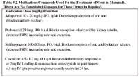
Table # 2. Medications Commonly Used for the Treatment of Gout in Mammals. There Are No Established Dosages for These Drugs in Reptiles5.
3.2 mg/dl (0.8 mmol/L). Correction may be achieved by careful slow intravenous infusion of calcium to effect. Oral calcium supplements, such as Neo-Calglucon (Nutrobal) are useful for long term calcium therapy. Caution must be exercised concerning the use of exogenous vitamin D3 for the potential to cause iatrogenic hypercalcemia and soft tissue mineralization may exist. The use of full spectrum light (i.e. Zoo med Reptisun 5.0 or Iguana Light) or preferably if possible direct unblocked exposure to natural sunlight should be provided to induce the natural endogenous synthesis of vitamin D3. The serum levels of calcium and phosphorus and the ratio should be monitored on a regular basis. Dehydration should be avoided at all costs by maintaining proper humidity levels, soaking the animals periodically, and adding water to food items. Anabolic steroids and vitamin B complex injections may be considered to stimulate the animals appetite and inhibit muscle catabolism. Nephrotoxic medications and any other undue stress (i.e., poor husbandry) to the animal should be avoided.
If acute renal disease or failure is not resolved, or chronic renal disease progresses untreated - then the outcome will most often be gout (visceral and/or articular). Acute gouty episodes may be treated symptomatically but widespread visceral gout is the result of end stage kidney disease and the prognosis is usually grave5 .

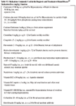
Table # 3. Medications Commonly Used in the Diagnosis and Treatment of Renal Disease2/7 Medication Dose (mg/kg) Function
As mentioned, gout is the end result of an inability of a reptile to properly excrete uric acid. Gout is defined as the deposition of uric acid crystals on visceral/parenchymous surfaces/organs and/or articular/joint surfaces. Gout is associated clinically with other disorders, especially those affecting water balance. Etiopathologic theories include inappropriate dietary nitrogen levels, renal disease and dehydration. Most likely any disturbance in renal excretion of uric acid in uricotelic species predisposes that individual to precipitation of urate crystals. Dietary management of gout has been tried in endotherms with disordered purine metabolism, but other supportive measures & medications are usually found to be more effective than changes in diet. However, dietary management may be warranted when animals are at risk for gout, or when a case is diagnosed early in its course. In animals, dietary management is achieved when rations are formulated with ingredients that are low in purines and that promote acidification.
Medical treatment of gout in human medicine is three-fold: Lower the serum uric acid level with antihyperuricemic drugs such as allopurinol; promote urate excretion with uricosuric drugs such as probenecid; and manage acute gouty arthritis attacks with anti-inflammatory drugs such as colchicine and corticosteroids. The treatment goals for gout in reptiles is similar to those in humans, however very little research has been done surrounding the treatment of gout in reptiles, and it is therefore not known/documented for certain whether or not the medications used in humans will achieve the same desired effects in reptiles. Also, in human medicine, the dosages for these drugs have been well established. Little information has been published for their use in reptiles. Therefore, the dosages used for reptile patients have been calculated extrapolating from the human dosages. These drugs are not without side effects - and therefore caution must be exercised when using these medications5.

Table # 4. Examples of Foods Low & High in Purines & To Promote Acidification
In conclusion, as stated, kidney related diseases are a major cause of illness and death in captive reptiles and amphibians. I believe that improper captive husbandry and diet are the most common predisposing causes of chronic renal failure in these animals. Therefore it must be the veterinarians goal to properly educate our client in the appropriate husbandry of their particular species when that animal is first seen in order to lessen the potential for renal problems in the future.
References & Suggested Readings
Divers, S.J. (1997). Clinician's Approach to Renal Disease in Lizards. Proc. 4th ARAV Conference, Houston, TX. p. 5 - 11.
Holz, P., et al. (1994) The Renal Portal System and Its Effect on Drug Kinetics. Proc. 1st ARAV Conference, Pittburgh, PA. p. 95 - 96.
Swenson, M.J. (ed.)(1984) Dukes' Physiology of Domestic Animals. Tenth Edition. Cornell University Press, Ithaca, NY. p. 474.
Mader, D.R. (1996). Reptile Medicine and Surgery. Saunders Co., Phil., PA. p. 374 - 379.
Frye, F.L. 1991. Biomedical and Surgical Aspects of captive Reptile Husbandry, 2nd edition, vol. 1. Krieger Publishing Co., Malabar, FL.
Jacobsen, E.R. (1988). Exotic Animals, Contemporary Issues in Small Animal Practice; Jacobsen ER, Kollias GV (eds). Churchill Livingstone, New York, NY. Chapt. 3, p. 35 - 48.
Bruederle, J.A. (1998). CVMA Exotics Formulary. Chicago, IL. p. 16 - 29. & Carpenter, J.W., Mashima, T.Y. and Rupiper, D.J. 1996. Exotic Animal Formulary, 1st edition.Greystone Publications. Manhatten, KS.