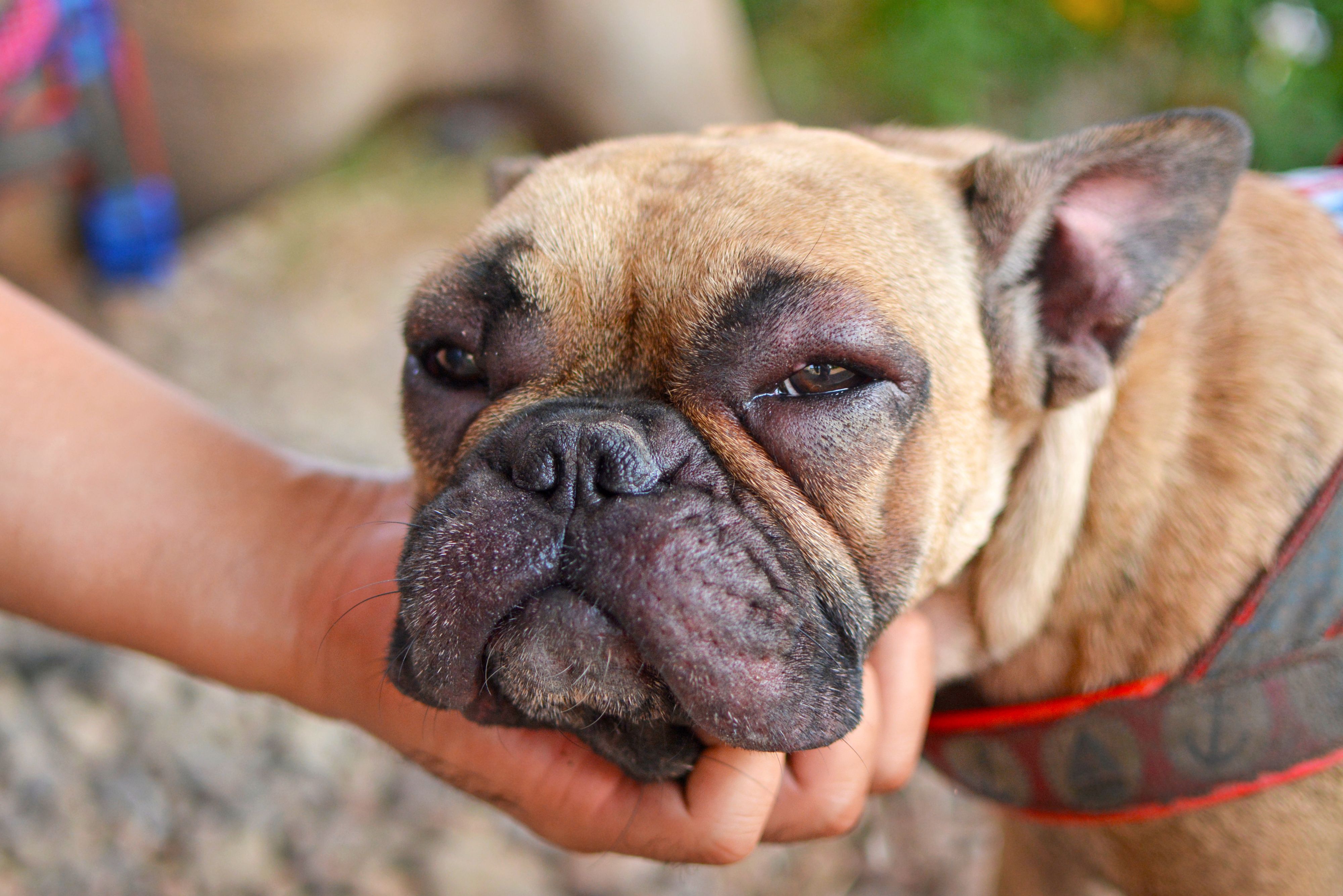
Approaching canine atopic dermatitis
Andrew Rosenberg, DVM, DACVD, detailed the ins and outs of canine atopic dermatitis, plus how to approach a patient presenting with this condition.
According to Andrew Rosenberg, DVM, DACVD, canine atopic dermatitis is a genetically predisposed inflammatory and pruritic allergic skin disease caused by environmental allergens with characteristic clinical features. It is the second most common allergic skin disease in dogs, following flea allergy dermatitis.1
At the 2021 New York Vet show in New York, New York, Rosenberg described the basics of canine atopic dermatitis and he shared insight on approaching a patient with this condition.
Atopic dermatitis basics
The primary clinical signs of atopic dermatitis include both erythema and pruritus and the secondary signs include excoriations and infections, said Rosenberg. It’s equally important to note that there are no pathognomonic signs and other skin diseases must be ruled out before making the diagnosis. “It’s not just an itchy skin disease, it’s also an inflamed skin disease,” he reminded.
The typical age of onset for atopic dermatitis is between 6 months to 3 years of age, and there is an increased risk in acquiring the condition for certain breeds such as Golden and Labrador retrievers, Pit Bull terriers, pugs, boxers, and German shepherds.2 Rosenberg revealed that the most common skin areas affected are the pinna, axilla, front paws, hind paws, lips, and the paranal.
He then detailed the mechanism of itch and inflammation surrounding canine atopic dermatitis. “Atopic individuals have a defective skin barrier…once this is defective, allergens can then absorb through the defective skin and allergens that interact with immune cells, those immune cells release a variety of itch chemicals…that gives the sensation of pruritus. A lot of other chemicals are being released by this allergic reaction many of which are very, very inflammatory.”
“That causes more inflammation on the skin and degrades that barrier function even more and then you get even more increased penetration of allergens…and secondary infections and bacteria,” Rosenberg continued.
Tips for approaching an atopic patient
Perform a cytology test or culture test
Rosenberg emphasized to begin by completing a cytology test when confronted with an atopic patient or completing a culture test if necessary.
“I really can’t stress enough to do your cytology’s, that should be the first thing you really are doing when you’re presented with an atopic dog,” he noted.
"Don’t be afraid to culture [a] skin infection, it’s never wrong to do so. If you see raw bacteria, you should be culturing and if there’s a bacterial infection that’s not resolving with antimicrobial therapies you should be culturing,” Rosenberg added.
Identify the underlying allergy
It is also key to determine which environmental allergy is affecting the patient so the client can limit the dog’s exposure to this allergen if possible. “It would be great if we could get to the root cause of what the dog is allergic to and manage that,” Rosenberg told attendees. Among the common allergens that cause a dog to develop atopy are various pollens, grasses, dander, insect proteins, and house dust.3
Treat the secondary infection
Once the pet is diagnosed and it is determined if it is a yeast or bacterial infection, treating the secondary infection is vital as this can significantly reduce pruritus, according to Rosenberg. Be sure to utilize the appropriate treatment method depending on your patient’s unique needs.
“My principles for treating secondary infections…we’re getting away as much as we can from systemic antibiotics with all the resistance…so if there’s [a] focal infection, I encourage you to use more topical therapies. For widespread infection, we still use antibiotics,” Rosenberg implored.
“I always recommend [prescribing an antibiotic for] at least 3 weeks, many times 4 weeks and the general rule is to treat with an antibiotic for 1 week beyond a clinical cure so you treat until everything looks normal and then add on a week,” he said. “I always recommend rechecking these patients before stopping [the] antibiotic."
References
- Elfenbein H. Atopic dermatitis in dogs: Causes, symptoms, and treatment. PetMD. February 13, 2020. Accessed November 8, 2021. https://www.petmd.com/dog/conditions/skin/c_dg_atopic_dermatitis
- Miller WH, Griffin CE, Campbell KL, eds. Hypersensitivity Disorders. In: Muller and Kirk’s small animal dermatology 7th ed. St. Louis, MO: Elsevier; 2013:372.
- The most common dog allergens and how to avoid them. Richell. September 20, 2019. Accessed November 8, 2021. https://www.richellusa.com/the-most-common-dog-allergens-and-how-to-avoid-them

