Babesiosis (Proceedings)
Canine babesiosis was first described in South Africa in the late 1800's and was originally presumed to be a "biliary form" of canine distemper. Most researchers assume that this was actually a case of babesiosis caused by Babesia canis rossi.
History
Canine babesiosis was first described in South Africa in the late 1800's and was originally presumed to be a "biliary form" of canine distemper. Most researchers assume that this was actually a case of babesiosis caused by Babesia canis rossi. The first report of canine babesiosis in the United States was made in 1934 and is presumed to have been caused by Babesia canis vogeli. The first report a small Babesia infection in a dog was in 1968. The first case of endemic babesiosis caused by a small piroplasm was in 1979. Since 1991 the number of cases of babesiosis caused by small Babesia spp. in the US has increased dramatically.
Classification
Historically Babesia spp. have been named/distinguished based on the vertebrate host infected and on the size of the red blood cell stage of the parasite. More recent molecular investigations have determined that this approach can be misleading, and it is now well accepted that specific DNA testing is the most accurate way to characterize the organisms causing Babesia infections. The majority of cases of canine babesiosis caused by large Babesia spp. in the US have been due to B. canis vogeli. However, molecular testing has identified a novel large Babesia spp. and has not been performed on most cases babesiosis. Therefore it is possible that other existing B. canis subspecies or novel large Babesia spp. that can infect dogs exist in the US. The majority of cases of canine babesiosis caused by small Babesia spp. in the US have been due to B. gibsoni (Asian genotype). At the time of this writing, there are at least nine genetically unique piroplasma that have been identified in the blood of dogs
Clinical signs and laboratory abnormalities
The following observations will pertain primarily to babesiosis in the United States. Lethargy, weakness, anorexia, pale mucous membranes and poor body condition are the most common clinical signs that cause owners to bring their pet to the hospital. Fever, lymphadenomegaly, splenomegaly, jaundice and pigmenturia are less commonly identified signs. Despite severe thrombocytopenia in some cases, petechiae and ecchymoses seem to be rare with babesiosis. Other animals are brought in for testing due to increased owner awareness and pre-breeding screening tests. It is very important to remember that many chronically infected dogs have no detectable physical abnormalities. Babesia infection is sometimes recognized as an incidental finding or is only recognized after immune suppressive therapy or splenectomy. Thrombocytopenia, hyperglobulinemia and anemia are the most common laboratory abnormalities detected. The thrombocytopenia can range from mild to severe, and is absent in some cases. The degree of anemia is variable and may be absent. Some cases present with a PCV of < 10% and others present with a PCV >50%. Most cases have a regenerative anemia at the time of presentation. Hypoalbuminemia, hyperbilirubinemia and pigmenturia are less commonly identified laboratory abnormalities.
Risk factors/transmission
Breed
Greyhounds are more likely to test positive for B. canis than other breeds. American Pit Bull Terriers are more likely to test positive for B. gibsoni than other breeds.
Ticks
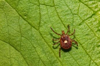
The presence of ticks at the time of presentation or even a history of tick attachment do not appear to be major risk factors for B. gibsoni infections in the US. The primary tick vectors for B. gibsoni, Haemaphysalis spp., have not been identified in the US. Rhipicephalus sanguineus is frequently referenced as a competent vector for B. gibsoni, but definitive transmission studies have yet to be performed. The tick vector for B. canis, R. sanguineus, is very common in the US.
Fights
Having been in a fight with an APBT is a risk for B.gibsoni infection.
Recent blood transfusion
Recent blood transfusion has been associated with infection in some dogs.
Kennels
Kennels in which one B. gibsoni infected dog is identified usually have a high prevalence of infection (10-50%). Racing Greyhounds from kennels have a higher prevalence of B. canis infection than pet greyhounds.
Breeding
Transplacental or perinatal transmission is very likely, but yet to be confirmed in a controlled setting.
Splenectomy/chemotherapy
A novel species of Babesia has been identified as a cause of thrombocytopenia, anemia, pigmenturia and fever in dogs that have undergone a splenectomy and/or are receiving chemotherapy.
Diagnostic testing
Microscopy
Organisms are most easily identified on 1000X. In house quick stains are usually adequate and in some cases superior for organism identification. Large piroplasms may migrate to the feathered edge during slide preparation. Use of capillary blood may increase organism recovery. Can result in both false positive and false negative tests. Microscopy is not useful for discriminating the genotype/species/sub-species. Cost: free to $86.00 (Antech path review)
Serology
Indirect immunofluorescent antibody (IFA) testing is the most commonly available test in the US. Difficult to interpret due to Lab to lab variation in methodology and different strains used for whole organism preps. The true sensitivity and specificity are not known. High titers usually correlate well with infection. However, IFA is not useful for the discrimination of genotype/species/sub-species. Other tests including ELISA's have been described, but are not commercially available. Cost: $18-50
Polymerase chain reaction
Amplifies a small fragment of Babesia DNA. Commercially available through several different sources. Not all PCR tests are the same. False positive and false negative tests are possible. Sensitive; one test detected B.gibsoni carriers 87% of the time and the sensitivity increased to 100% when two consecutive tests were performed. Only way to definitively identify the genotype/species/subspecies. Cost: $50-55
Other
Many (>50%) of cases are Coomb's test positive. Co-infection with Ehrlichia, Bartonella, Mycoplasma, Leishmania and/or Rickettsia have been identified in some cases, so testing for the presence of other infectious diseases is warranted.
Treatment
Note
Since the incidence of canine babesiosis is increasing in the US, specific anti-Babesial treatment is appropriate and indicated in dogs highly suspected of having babesiosis even without a definitive diagnosis.
Antibiotics
Antibiotics are not the treatment of choice, but are reasonable to consider while waiting for specific anti-protozoal therapy. Doxycycline will reduce clinical signs and is associated with decreased morbidity and mortality. The dose is 10mg/kg/day. Administered IV or PO once or twice daily. I prefer once daily dosing unless the patient is vomiting after the medications. Clindamycin has been used to treat babesiosis. The dose in a recent study was 25mg/kg PO BID for 14 days, and was associated with a reduction of clinical, hematological and biochemical abnormalities. It is important to remember that antibiotics alone have not been shown to clear infections. Administration of antibiotics alone without specific testing has resulted in a number of dogs being tentatively diagnosed with ehrlichiosis based on a partial or complete recovery. Cost: $10-50
Imidocarb diproprionate
Imidocarb diproprionate (Imizol®, Schering-Plough) is the only FDA approved drug for the treatment of canine babesiosis. Dose 6.6mg/kg IM once. Repeat dose in 2 weeks. I will usually re-dose in one week if the dog is not responding well. Likely to cure B. canis vogeli. Good to reduce morbidity, mortality and parasitemia in B. gibsoni (Asian genotype), but is not curative. Side-effects include pain at injection site and cholinergic effects (SLUD). Pre-treatment with atropine reduces cholinergic effects. Cost: $50-100
Atovaquone and azithromycin
An atovaquone and azithromycin drug combination is the only treatment shown to reduce B. gibsoni parasitemia below the limit of detection by a sensitive PCR test. Was effective in 83% of treated dogs in a controlled trial. Atovaquone is an anti-protozoal drug believed to inhibit the action of cytochrome B. It is available in two formulations in the US, Mepron® (GlaxoSmithKline) and Malarone® (GlaxoSmithKline). Mepron is a single drug formulation and is available in a liquid suspension. Malarone is a multi-drug combination containing atovaquone and proguanil hydrochloride. In our experience Mepron is more expensive, but is very well tolerated, while Malarone has been associated with severe GI side-effects. The atovaquone dose is 13.5mg/kg PO TID administered with a fatty meal for 10 consecutive days in combination with azithromycin. The azithromycin dose is 10mg/kg PO Q24 for 10 consecutive days. We have not used this combination to treat B. canis or B. gibsoni (USA/California genotype). Cost: $300-900
Diminazene aceturate
Not FDA approved. Dose 3-7mg/kg IM once. Diminazene is likely to be curative for B. canis. It does not cure B. gibsoni (Asian genotype), but reduces morbidity, mortality and parasitemia. Anecdotal evidence suggests that B. gibsoni infected dogs recover more quickly than those treated with imidocarb. Side-effects are similar to imidocarb. Mortality has been noted at doses greater than 10mg/kg. Cost: your veterinary license?
Supportive care
Intravenous fluids are indicated for hypovolemic patients. Whole-blood or Packed RBC transfusions (from screened donors of course) for dogs that are clinical for their anemia. We have also used a hemoglobin based oxygen carrying solution in some dogs with babesiosis.
Immune-suppressive drugs
The use of immune-suppressive drugs for the immune-mediated components of the disease (anemia and thrombocytopenia) is controversial. If a dog is stable (i.e., eating and drinking and does not require ICU), regardless of PCV, I will treat with anti-protozoal drugs alone. I have treated dogs with PCV's as low as 8% with imidocarb alone and they have done very well. If a dog is crashing despite anti-protozoal therapy, I will treat like an IMHA case with 2mg/kg/day of prednisone. I will taper the prednisone faster in these cases than I do with idiopathic IMHA cases. Cases that have been treated with long-term immune-suppressive drugs prior to specific anti-protozoal treatment tend to have a poor response to treatment.
Follow-up
Time to respond
Most dogs begin to improve within 3-7 days of specific anti-protozoal treatment with some dogs taking up to 14 days. Dogs that have been treated with long-term immune-suppressive drugs prior to specific anti-protozoal therapy tend to take longer to respond or fail to respond.
Recovery
Babesia canis infected dogs typically have a complete recovery after treatment. Treatment failures are less common with B. canis. Babesia gibsoni infected dogs treated with atovaquone and azithromycin combination therapy have not been reported to have relapse of clinical disease and some dogs have remained PCR negative for over three years. Babesia gibsoni infected dogs that remain persistently infected after specific therapy will typically fall into one of the following categories:
1. "Complete recovery" means there is resolution of anemia and no outward signs of disease even after stress or illness. Mild thrombocytopenia and hyperglobulinemia often persist.
2. "Partial recovery" means that outward clinical signs have resolved, but mild anemia (PCV 25-35%), thrombocytopenia and hyperglobulinemia persist.
3. "Complete or partial recovery, but relapse during stress" means that the outward clinical signs and possibly clinicopathological signs resolve, but during periods of stress (whelping, illness, fight, etc.) the dog becomes clinically ill again.
Follow-up testing
Serial blood counts are recommended until PCV and platelet count return to within normal range. In a laboratory setting splenectomy and/or sub-inoculation into a splenectomized dog are the gold-standards to document the clearance of infection. Serology is a poor short-term follow-up tool as titers may persist for months to years regardless of infection status. If a titer falls below the limit of detection, it is likely to correlate with the resolution of infection, if it was tested in the same laboratory with the same antigen preparation and operator. Microscopy and PCR are the tests that I recommend for post-treatment follow-up. Since the half-life of many of the treatment drugs is long, follow-up tests should be performed 60 days post-treatment. Since the sensitivity of PCR is not 100%, we recommend two consecutive negative tests.
Other considerations
Since confirmation of a complete cure cannot be confirmed without splenectomy or sub-inoculation of blood into a splenectomized dog, caution should be used when using treated animals for breeding etc.
References available from author upon request
Newsletter
From exam room tips to practice management insights, get trusted veterinary news delivered straight to your inbox—subscribe to dvm360.


