Canine leishmaniasis: Update in dogs (Proceedings)
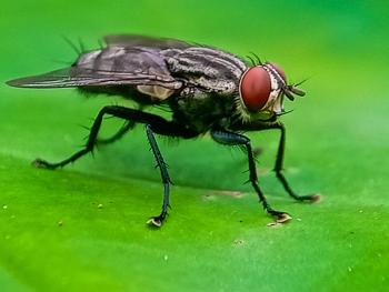
Sandfly vectors (not in US) transmit flagellated parasites into the skin of a host, where it often localizes in the cat. In dogs, there is invariably spread of the parasite throughout the body to most organs, although renal failure is the most common cause of death.
Overview
• The protozoan of the genus Leishmania causes two types of disease - cutaneous or visceral. Organ systems affected include the skin, hepatobiliary, spleen, kidneys, eyes, and joints, or develop hemorrhagic diathesis.
• Dogs with Leishmaniasis in the United States fall into 2 categories:
1. acquired the infection (usually caused by L. donovani infantum) overseas from around the Mediterranean basin, Portugal, and Spain, with sporadic cases arising from Switzerland, northern France, and the Netherlands. Dogs imported from endemic areas of South and Central American, and southern Mexico may be infected with L. donovani complex or L. braziliensis.
2. Endemic cases in Foxhounds populations (Feb 2000) in most states and even Canada. Thought not to be vector transmitted.
• Sandfly vectors (not in US) transmit flagellated parasites into the skin of a host, where it often localizes in the cat. In dogs, there is invariably spread of the parasite throughout the body to most organs, although renal failure is the most common cause of death. Incubation period: 1 mth - yrs.
Signalment
• Virtually all dogs develop visceral or systemic disease, but 90% of dogs have cutaneous involvement as well. There is no sex or breed predilection (except Foxhounds in US!).
• Cats develop cutaneous disease (rare) with no sex or breed predilection.
Signs
Visceral
• Historically, exercise intolerance, severe weight loss, anorexia are the main signs with diarrhea, vomiting, epistaxis and melena less common.
• Clinically, most dogs have lymphadenopathy, cutaneous lesions, and emaciation. Signs of renal failure (polyuria, polydipsia, vomiting) may be present. Neuralgia, polyarthritis, polymyositis, osteolytic lesions or proliferative periostitis are rare. Fever and splenomegaly occurs in about a third of dogs.
Cutaneous
• Hyperkeratosis is the most prominent finding (excessive epidermal scale with thickening, depigmentation and chapping of the muzzle and footpads). Hair coat is dry, brittle with hair loss. Intradermal nodules and ulcers occur in some dogs. Abnormally long or brittle nails are a specific finding in some dogs.
• Cats usually develop cutaneous nodules.
Differential DIAGNOSIS
• Differentiate visceral from systemic diseases including mycoses (blastomycosis, histoplasmosis), systemic lupus erythematosus, metastatic neoplasia, distemper, vasculitis.
• Cutaneous lesions need to be differentiated from other causes of hyperkeratosis including primary idiopathic seborrhea and nutritional dermatoses (vitamin A-responsive, zinc-responsive). Other causes (idiopathic nasodigital hyperkeratosis, lichenoid-psoriasiform dermatosis, epidermal dysplasia, and Schnauzer comedo syndrome) are rare and breed specific. Hyperkeratotic and nodular lesions caused by leishmaniasis are easily differentiated from other etiologies by finding organisms on skin biopsies.
Laboratory tests
• Hyperproteinemia with hyperglobulinemia (100% of cases) and hypoalbuminemia (95% of cases) is characteristic (differentiate from chronic ehrlichiosis and multiple myeloma).
• Proteinuria (85% of cases), high liver enzyme activity (55% of cases), thrombocytopenia (50% of cases), azotemia (45% of cases), and leukopenia with lymphopenia (20% of cases) are other findings.
• Coomb's test, antinuclear antibody test, and lupus erythematosus cell test may be positive.
• Serologic diagnosis is available – fluorescent antibody (Communicable Disease Center - CDC), or ELISA from other diagnostic laboratories (Cornell, North Carolina).
• Culture of skin, splenic, bone marrow, or lymph node biopsies or aspirates by the CDC although this takes a long time and is not practical.
• Cytologic and histopathologic identification of intracellular organisms in the above biopsies or aspirate specimens is the definitive test of choice.
• Cytologic examination of the lymph nodes and bone marrow is probably not as efficient as culture of the lymph nodes or bone marrow.
• PCR of bone marrow tissue particularly, but also lymph nodes is effective especially in heavily infected animals.
• A combination of diagnostic techniques (cytology, serology etc) is often needed to make a definitive diagnosis.
Pathologic findings
• Cell infiltration (mainly histiocytes and macrophages) and characteristic intracellular amastigote forms can be identified in many tissues including skin, lymph nodes, liver, spleen, and kidney.
• May use immunohistochemistry to differentiate from Trypanosoma cruzi amastigotes if in doubt.
• Mucosal ulcerations in the stomach, intestine and colon can be seen occasionally.
Treatment
• As prognosis is very poor for emaciated chronically infected animals, consider euthanasia.
• Advise owner of potential zoonotic transmission of organisms in lesions to humans.
• Treat on an outpatient basis. Provide a high good quality protein diet or a diet appropriate for renal insufficiency if present.
• Single dermal nodule lesions in cats are best surgically removed.
• Inform owners organism will never be eliminated and relapse is inevitable requiring re-treatment.
Drugs
• The pentavalent antimonials have been the mainstay of treatment in dogs in Europe but are difficult to obtain the USA – need to obtain from the CDC. Two main drugs in this class:
1. Sodium stibogluconate (Pentostam®; Wellcome Foundation Ltd, United Kingdom). Requires daily injection and has severe side effects. Resistance has been reported in France, Spain, and Italy. Dose: 30-50 mg/kg IV or SC, q24h, for 3 to 4 wks.
2. Meglumine antimonite (Glucantime®; Merial, Lyon, France). Less side-effects than Pentostam®. Dose: 100 mg/kg, SC, q24h, for 3 to 4 weeks (lower dose in ill dogs).
• In the USA, it is recommended to begin with Allopurinol. The organism metabolizes allopurinol to produce an analogue of inosine which is incorporated into the leishmanial RNA resulting in faulty parasite multiplication. It is efficacious and non-toxic when used as a maintenance drug. Clinical remission is often achieved when used alone. Doses vary widely but 20 mg/kg, PO, q12h results in rapid clinical remission – relapses are common if drug is stopped; complete cures are rare but survival occurs in about 80% of cases over 4 years if renal insufficiency is not present when treatment is initiated. Another effective reported dose: 15 mg/kg, PO, q12h.
• Amphotericin B. In the lipid emulsion or liposomal form, is relatively non-nephrotoxic and is effective against the organism, although not proven so far to be superior to allopurinol (more costly and toxic also.
• Treat renal insufficiency before giving antimonial drugs as prognosis is dependent on renal function at the onset of treatment.
• Treatment efficacy: best monitored by clinical improvement and presence of organisms in biopsy
• Relapses occur a few months to a year after therapy, so recheck at least every 2 months after end of treatment.
• Prognosis for a cure is very guarded, but therapy gives the dogs some quality of life.
References
1. Lindsey DS, Zajac AM, Barr SC. Leishmaniasis in American Foxhounds: An emerging zoonosis? Comp Cont Ed Pract Vet 24:304-312, 2002.
2. Baneth G. Leishmaniasis. In: Greene CE, ed. Infectious diseases of the dog and cat. Philadelphia: WB Saunders, 2006: 685-698.
3. Rosypal AC, Troy GC, Duncan RB et al. Utility of diagnostic tests used in diagnosis of infection in dogs experimentally inoculated with a North American isolate of Leishmania infantum. J Vet Intern Med 19:802-809, 2005.
4. Petersen CA and Barr SC. Canine Leishmaniasis in North America: Emerging or newly recognized. Vet Clin Nth Am: Small An Pract; Small An Parasites: Biology and Control. 2009;39:1065-1074
5. Schantz PM, Steurer FJ, Duprey ZH, et al. Autochthonous visceral leishmaniasis in dogs in North America. J Am Vet Med Assoc 8:1316-1322, 2005.
Newsletter
From exam room tips to practice management insights, get trusted veterinary news delivered straight to your inbox—subscribe to dvm360.


