Caring for cats with cardiomyopathies
Veterinary cardiologist Dr. Meg Sleeper outlines everything you need to know about the three types of primary feline cardiomyopathy you may encounter in your practice.
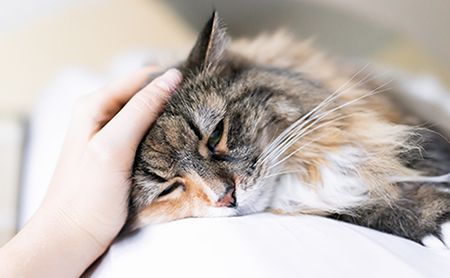
Kristina /stock.adobe.com

While cardiomyopathies have long been the most common cardiac diseases found in cats, they still can be difficult to detect and treat, says Meg Sleeper, VMD, DACVIM (cardiology), professor of cardiology at the University of Florida College of Veterinary Medicine in Gainesville.
“Detecting cardiomyopathies in cats is difficult because feline cardiac disease is so unpredictable,” Dr. Sleeper said in a recent conversation with dvm360 magazine. A full 50% of cats with heart disease will not have a detectable murmur, she noted. And of those that do, only about 50% will have echocardiographic evidence of heart disease.
Types of cardiomyopathy
Hypertrophic cardiomyopathy. Hypertrophic cardiomyopathy (HCM) is the most common primary cardiomyopathy in cats. Cats with HCM have a defect in their cardiac myocytes, causing the muscle cells to be in disarray rather than in a regular, organized pattern. HCM is characterized by concentric hypertrophy and fibrosis of the left ventricle. This causes stiffening of the ventricle, which prevents relaxation and impairs ventricular filling during diastole.
In ragdoll and Maine coon cats, the disease is caused by an autosomal dominant mutation with incomplete penetrance. This means that all cats with the mutation will have some form of HCM, even if they are heterozygous for the mutation. Dr. Sleeper added that other, undiscovered genetic mutations are likely the cause of HCM seen in other breeds, including domestic shorthair cats.
Manifestation of HCM can vary widely from patient to patient, with variables including age, areas of the heart affected, presence of dynamic outflow obstruction, disease severity and clinical presentation:
Age:Age of onset ranges from 6 months to 16 years (mean, 6 years).
Areas affected: Some cats will have widespread concentric hypertrophy, while others will have only a focal area of hypertrophy. “This underscores the need to look at the entire ventricle during echocardiography,” Dr. Sleeper said.
Dynamic outflow obstruction: Some cats will experience systolic anterior motion (SAM) of the mitral valve, in which the mitral valve is drawn into the left ventricular outflow tract during systole. Moderate or severe SAM increases left ventricular systolic pressure, which exacerbates left ventricular hypertrophy, Dr. Sleeper said. “This creates a vicious cycle of hypertrophy and the potential for worsened diastolic function,” she added.
Disease severity: Some cats may have very mild, slowly progressive disease, never becoming symptomatic, while others will develop more severe disease that may lead to arterial thromboembolism, congestive heart failure (CHF) or sudden death. “About half of cats that go into CHF have some precipitating event, such as a dental or other stressful procedure,” Dr. Sleeper said.
Clinical presentation: Although many cats with HCM are asymptomatic, 36% to 72% have a systolic murmur, 33% have a gallop rhythm, 35% have dyspnea, 4% have syncope and others will have nonspecific signs, such as lethargy, hyporexia or vomiting.
Restrictive cardiomyopathy. Like HCM, restrictive cardiomyopathy affects diastolic function, Dr. Sleeper said. The disease is characterized by focal, regional fibrosis of the left ventricular myocardium, causing stiffening of the left ventricle and inhibiting passive filling during diastole. Unlike HCM, restrictive cardiomyopathy does not cause ventricular wall thickening.
Dilated cardiomyopathy. Dilated cardiomyopathy (DCM) is characterized by a dilated heart with thin walls and decreased systolic function. The disease was once common in cats but has become quite rare since the association between DCM and insufficient dietary taurine was discovered. The disease is more common in Abyssinian, Burmese and Siamese cats than in other breeds.
Diagnostic testing
The variability in signalment and presentation of cats with cardiomyopathy can make the condition difficult to recognize and diagnose. While echocardiography is needed to diagnose and differentiate between the cardiomyopathies, other diagnostic tests play an important role.
Lab work. If a primary cardiomyopathy is suspected, diseases that can cause secondary hypertrophy should always be ruled out, said Dr. Sleeper. “Cats with CHF or suspected HCM should have comprehensive lab work performed, including a complete blood count, serum chemistry, total thyroxine level and urinalysis,” she said. This is especially true for middle-aged and older cats, which are more likely to have renal disease or hyperthyroidism. Renal disease can contribute to hypertension, and it's important to assess renal function before initiating treatment for CHF. Assessing total thyroxine concentration is essential, Dr. Sleeper said, because hyperthyroidism is a secondary cause of concentric hypertrophy.
Acromegaly is a less common cause of secondary HCM. While serum growth hormone levels can be measured to test for acromegaly, checking growth hormone levels usually is not necessary because acromegaly often is evident on physical exam, said Dr. Sleeper.
Electrocardiography. Some cats with cardiomyopathy may have changes on an electrocardiogram (ECG), while others will have a completely normal ECG. The most common changes are tall R waves (indicating left ventricular hypertrophy), tall P waves (indicating right atrial enlargement) and arrhythmias. “Electrocardiography is not the most sensitive test,” Dr. Sleeper said, “but it is an important test in cats that have a history of arrhythmia or syncopal episodes.”
Radiography. Thoracic radiographs in cats with cardiomyopathy may look normal, or they may show left atrial enlargement or generalized cardiomegaly. Radiographs also can show evidence of CHF. Serial vertebral heart scoring is an inexpensive, objective method to monitor heart size and disease progression when client finances are limited, Dr. Sleeper said. Because measurements of both the left atrium and the left ventricle are captured with a vertebral heart score, the score will change with enlargement of either chamber.
Cardiac pro-brain natriuretic peptide (pro-BNP). BNP is a vasodilatory and natriuretic protein that is released from the ventricular myocardium in response to physical stress (i.e. increased stretching, increased wall tension or ischemia). BNP levels can be useful in detecting cardiomyopathy in cats. There are very few cats with a pro-BNP level higher than 100 pmol/L that do not have HCM, but the test can miss those with mild to moderate disease. In other words, at higher than 100 pmol/L the test will have very few false positives but can have false negatives. “The pro-BNP level will often rise prior to radiographic evidence of cardiac enlargement in the cat,” Dr. Sleeper said.
Echocardiography. As noted earlier, many diagnostic tests can be suggestive of cardiomyopathy, but echocardiography is needed to differentiate between the types of cardiomyopathies. Echocardiography is used to measure ventricular wall thickness, identify myocardial fibrosis, assess for SAM of the mitral valve, measure pressure gradients, measure left atrial size and assess for atrial blood stasis and thrombus formation. In cats, the diameter of the left atrium should approximately equal the diameter of the aorta. The magnitude of left atrial enlargement is a very important prognostic indicator, said Dr. Sleeper, who uses this measurement in determining when to initiate treatment.
Treatment and monitoring
Treatment of cats with HCM and restrictive cardiomyopathy can be controversial, Dr. Sleeper said, adding that “the best medicine for some cats may be no treatment at all.” Because some asymptomatic cats will never develop severe disease, it's difficult to know when to begin treatment. Factors to consider before starting treatment include the presence of left atrial dilation, a history of previous thromboembolism, severity of SAM of the mitral valve, tachycardia, severity of left ventricular hypertrophy, and client and patient motivation and ability to administer medications long term. If the client and patient can tolerate treatment, Dr. Sleeper said she generally will start treatment if a consistent tachycardia or arrhythmia is present, if there is severe dynamic stenosis or if there is evidence of significant left atrial enlargement.
Atenolol and diltiazem are the most commonly used drugs in asymptomatic cats with HCM. Atenolol is more useful than diltiazem in treating tachycardia and reducing SAM of the mitral valve. Angiotensin-converting enzyme (ACE) inhibitors also have been used in asymptomatic cats with HCM. Clinical evidence from placebo-controlled, blinded clinical studies indicate that early use of ACE inhibitors or diuretics in cats with asymptomatic HCM is not warranted, Dr. Sleeper said.
Prophylactic anticoagulant therapy in asymptomatic cats with HCM also is controversial, according to Dr. Sleeper, and generally is not needed in cats with HCM and normal left atrial size. Cats with echocardiographic evidence of spontaneous contrast (red blood cell aggregation), intracardiac thrombus, a history of previous thromboembolism, and moderate to severe left atrial dilation (left atrial-to-aortic ratio ≥1.9 and/or evidence of atrial blood stasis) should be placed on anticoagulant therapy. Clopidogrel, aspirin and low-molecular-weight heparin are used as anticoagulants in cats. Although clopidogrel is superior to aspirin as an anticoagulant, it is extremely bitter and giving the medication can become a quality of life issue for patients and their families, Dr. Sleeper said, adding that aspirin may be preferable in these cases.
Treatment for cats with DCM is less controversial. “DCM is treated with taurine supplementation, pimobendan and an ACE inhibitor,” said Dr. Sleeper, who sees a handful of feline DCM cases each year.
Furosemide is the most effective and lifesaving treatment for cats with CHF, she said. Depending on the severity of heart failure, furosemide can be given orally at 1 to 2 mg/kg every eight to 24 hours as outpatient therapy. The lowest effective dose should be used, and a higher initial dose often can be tapered rapidly, based on respiratory rate and effort as well as evaluation of thoracic radiographs. In acute heart failure, parenteral furosemide is given at a dose of 1 to 2 mg/kg every one to four hours, tapering the dose and frequency once respiratory effort normalizes and the respiratory rate decreases to 50 bpm or less.
Oxygen therapy with 60% to 70% fraction of inspired oxygen also can be used in patients with acute heart failure but should be decreased to 50% or less within 12 hours to avoid barotrauma secondary to high inspiratory oxygen concentration, Dr. Sleeper cautioned. Cats in acute heart failure at high risk for arterial thromboembolism are started on prophylactic anticoagulants while hospitalized.
Pimobendan may also be used in with patients acute heart disease, ideally based on echocardiographic findings. Negative inotropes, such as atenolol and diltiazem, generally are not indicated in acute cases, unless the patient has a hemodynamically significant tachyarrhythmia. Beta-blockers, such as atenolol, should be avoided until heart failure has been stabilized. ACE inhibitors also are not used in emergent cases, said Dr. Sleeper, adding that they can be useful as an adjunct treatment once the cat is stable at home and well hydrated.
Middle-aged to older cats should be monitored periodically for development of hypertension and hyperthyroidism, as these conditions can accelerate cardiac disease progression. Asymptomatic cats with HCM should have an echocardiogram every 6 to 12 months, depending on the severity and progression of their disease. Cats with significant left atrial enlargement should have radiographs every 3 to 6 months to evaluate for early heart failure. Those that are already in heart failure should have repeat thoracic radiographs and renal panels performed to monitor clinical response and to guide medical therapy, Dr. Sleeper advised, adding that thoracic ultrasound can be done to evaluate pleural effusion and to guide thoracentesis.
Prognosis
Cats with asymptomatic and mild disease have a good prognosis, and may live for years without problems, Dr. Sleeper said, but the prognosis worsens once cats develop CHF and arterial thromboembolism. Survival times in cats with CHF range from 92 to 654 days. Cats with HCM and arterial thromboembolism have an average survival time ranging from 61 to 184 days.
