|Articles|December 1, 2005
Conservative treatment of atlantoaxial subluxation in canine patients
In a recent article [ME Havig et al.: Evaluation of non-surgical treatment of atlantoaxial subluxation in dogs: 19 cases (1992-2001) in JAVMA, Vol. 227, No. 2, July 15, 2005], it was suggested that non-surgical treatment (neck-brace application) of acute atlantoaxial subluxation (AAS) carries a good long-term outcome in about 62 percent of the cases. The success rate of surgical treatment was cited as a 61-91 percent in the same paper.
Advertisement
In a recent article [ME Havig et al.: Evaluation of non-surgical treatment of atlantoaxial subluxation in dogs: 19 cases (1992-2001) in JAVMA, Vol. 227, No. 2, July 15, 2005], it was suggested that non-surgical treatment (neck-brace application) of acute atlantoaxial subluxation (AAS) carries a good long-term outcome in about 62 percent of the cases. The success rate of surgical treatment was cited as a 61-91 percent in the same paper.
Photos 1-3: Example of a neck brace with a dorsal splint and soft-padded bandage. The brace had to be removed and applied more loosely a few minutes after the first application due to cyanosis.
In the article, patients with clinical signs for less then 30 days were defined as an acute subluxation. Dogs with chronic subluxation (signs of AAS for more than 30 days) had significantly worse prognosis when compared to the acute cases. Interestingly, the neurologic condition at admission, the radiographic appearance of the dens and the clinical history did not affect the clinical outcome.
Advertisement
Diagnosing AAS can be challenging. Small and toy-breed dogs are predisposed due to their several anatomic abnormalities (such as odontoid process agenesis, hypoplasia or dorsal angulation of the dens). The recommended radiographic projections for the visualization of the dens are the flexed lateral and the "open-mouth" views under general anesthesia. Unfortunately, these positions can subluxate the unstable joint further, causing additional spinal cord compression. (JP Morgan, CS Bailey: Exercises in veterinary radiology: spinal disease. Venture Press, 1st ed., 2000).
The clinical signs at presentation may vary from stiffness of the neck to tetraparesis. Severe or minor trauma can precede the initial signs.
The theory behind the non-surgical approach is an attempt to stabilize the cervical vertebral column in a mildly extended position, allowing the body to build a fibrous connective tissue around the atlanto-axial joint over four to 15 weeks. There are several ways to apply a neck brace using fiberglass, splint or soft-padded bandage. The splints in this study were applied to the dorsal, ventral or both sides of the bandage. The ventral splint extended from the rostral mandible to the xiphoid, the dorsal splints from the bony orbit to the last thoracic vertebra.
Complications can occur following splint application. The most severe complications are cyanosis, dyspnea and death secondary to a very tight bandage. It is advisable that the patient be closely observed after splint application during the first day. Periodic rechecks and bandage changes are advisable. If the bandage becomes wet, it might tighten and cause complications. Clients must be prepared for the chance of relapse and thus possible surgical stabilization when the splint is removed. Crate rest is recommended for several weeks.
By Beatrix Nanai DVM Dr. Nanai is a resident of the European College of Veterinary Neurology/Neurosurgery at the Animal Emergency andReferral Center in Fort Pierce, Fla.
By Ronald Lyman DVM, Dipl. ACVIM Dr. Lyman is a graduate of The Ohio State University College of Veterinary Medicine. He completed a formal internship at the Animal Medical Center in New York City. Lyman is a co-author of chapters in the 2000 editions of Kirk's Current Veterinary Therapy XIII and Quick Reference to Veterinary Medicine.
Newsletter
From exam room tips to practice management insights, get trusted veterinary news delivered straight to your inbox—subscribe to dvm360.
Advertisement
Related Content
Advertisement
Advertisement
Advertisement
Trending on dvm360
1
Q&A: What the 2023 ACVIM update means for leptospirosis vaccination
2
Nonpharmacologic ways of managing pain and separation anxiety in dogs
3
Clinic center: Schwarzman Animal Medical Center completes $125 million renovation, and other updates
4
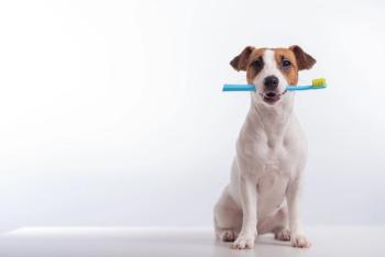
Tarter control toothpaste receives VOHC Seal of Acceptance
5