
|Articles|May 1, 2011
Correcting ectopic ureters in juvenile dogs
Cystoscopic-guided laser ablation is a minimally invasive alternative to surgery for these congenital anomalies.
Advertisement
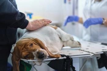
Ectopic ureters are a congenital anomaly of the urinary system, in which the ureteral orifice is inappropriately positioned caudal to the urinary bladder. This is the most common cause of urinary incontinence in juvenile female dogs. See how the use of cystoscopic-guided laser ablation provides a minimally invasive alternative to surgery in cases with intramural ectopic ureters, as was seen in this Golden Retriever puppy.
Initial findings
- Signalment: 4-month-old intact female Golden Retriever
- Presenting complaint: Owner is unable to housetrain; urine staining around hind end
- Pertinent history: Always wet around back end; bad-smelling urine; urine culture results positive for Escherichia coli that is sensitive to amoxicillin-clavulanic acid; with treatment, odor resolved, but incontinence persisted
- Medications: Amoxicillin-clavulanic acid (15 mg/kg orally b.i.d.), phenylpropanolamine (1.5 mg/kg orally t.i.d.)
- Physical examination findings: Bright, alert, good body condition; urine staining around fur of tail, vulva and medial aspect of both hindlimbs; slightly recessed vulva; normal neurologic examination with good anal tone. The remainder of the examination was within normal limits. Rectal examination palpated a wide urethra with evidence of the bladder neck at the level of the pubis.
Diagnostic evaluation
- Blood pressure: 130 mm Hg systolic
- Complete blood count: Slight normochromic normocytic nonregenerative anemia of 33 percent
- Serum chemistry profile: BUN 13, creatinine 0.4, phosphorus 12
- Urinalysis: USG 1.021, no white blood cells, no red blood cells/hpf, no crystals, no bacteria, pH 6.5
- Urine culture: Negative (while still receiving amoxicillin-clavulanic acid)
- Abdominal radiography: Within normal limits
- Abdominal ultrasonography: Loss of architecture to both kidneys; minimal pyelectasia (2.5 mm) bilaterally; empty bladder, unable to perform cystocentesis
• Cystoscopy: See Figure 1
Figure 1: Endoscopic images of a dog with ectopic ureters. The dog is in dorsal recumbency during a cystourethroscopy. A) The left ectopic ureteral opening is visualized inside the urethral lumen (yellow asterisk). B) An open-ended ureteral catheter is placed inside the ectopic ureteral lumen (black arrow). C) A diode laser (red arrow) is cutting the medial ureteral wall over the ureteral catheter (black arrow) to advance up the neo-ureteral orifi ce to the bladder lumen. D) The neo-ureteral orifi ce is now inside the urinary bladder lumen (yellow asterisk). A guidewire (black arrow) is still inside the ureteral lumen.
Advertisement
• Retrograde ureteropyelography: See Figure 2
Figure 2: A fl uoroscopic image of a dog during retrograde ureteropyelography and concurrent cystourethrography. The bladder is fi lled with contrast material. The rigid cystoscopy is at the level of the bladder trigone, and a guidewire is inside the ureteral lumen coursing through the intramural tunnel and then transitions extramurally beyond the bladder trigone.
• Vaginoscopy: See Figure 3
Figure 3 (far right photo): Endoscopic images with the dog in dorsal recumbency after the cystoscopic-guided laser ablation procedure. The top image shows a thick vaginal band (persistent paramesonephric remnant) pulling the urethral orifi ce open. This band splits the vaginal opening into two compartments. The middle image is the remnant of the vaginal band after it is laser-ablated with a diode laser. This band went all the way back to the cervix and was completely cut down with the laser to the level of the cervix seen here. The bottom image is the vaginal (bottom) and urethral orifi ce (top) after thepersistent paramesonephric remnant is lasered open showing an open vagina.
Problem list
- Urinary incontinence
- History of urinary tract infection
- Recessed vulva
- Bladder neck caudally displaced
Presumptive diagnosis
- Possible ectopic ureter or ureters
- Urethral sphincter mechanism incompetence (USMI)
- Short urethral syndrome
Diagnoses
- Bilateral intramural ureteral ectopia
- Bilateral hydroureter and hydronephrosis
- Short urethra syndrome with a hypoplastic bladder
- Persistent paramesonephric remnant that extended back to the cervix (vaginal septum)
Treatment decisions
During the diagnostic cystoscopy, a diode laser was used to perform a cystoscopic-guided laser ablation of the intramural bilateral ectopic ureters (CLA-EU). The right ureter was displaced in the mid urethra and the left was in the proximal urethra. Both were intramurally tunneling from the bladder into the urethral lumen where the opening was located. This was diagnosed based on the retrograde ureteropyelogram in which the ureters and renal pelvis were seen to be dilated. Once the CLA-EU was complete, the laser was used to ablate the vaginal remnant and create a normal vagina to try to prevent urine pooling and vaginitis in the future.
Outcome
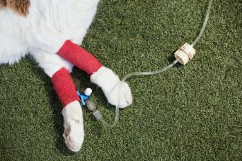
The patient was discharged the same afternoon as the cystoscopic procedure. The amoxicillin-clavulanic acid was continued for two weeks, and tramadol was prescribed (3 mg/kg orally t.i.d.) for two days as needed. During the next four weeks, the dog was completely continent. Urine culture results at four weeks were negative. The dog's renal pelvis size was reassessed six weeks after the procedure, and there was no evidence of renal pelvic dilation of hydroureter bilaterally.
At 7 months of age, the dog started having urine spotting at night, at which time the phenylpropanolamine was started at 1.5 mg/kg orally before bed. This made the dog 100 percent continent. At 2 years old, the dog has no evidence of urinary incontinence.
Discussion
The embryologic foundation of ectopic ureters is thought to result from the abnormal differentiation of mesonephric and metanephric duct systems, resulting in inappropriate ureteral tube termination and malposition of the ureteral orifice.
Although ectopic ureters have been reported in male and female dogs, as well as both purebred and mixed-breed dogs, it seems to occur with greater frequency in female dogs and in certain breeds, including Siberian Huskies, Newfoundlands, Labrador retrievers, Golden Retrievers, terriers and poodles. The most common clinical finding is constant or intermittent urinary leaking since birth or weaning, though many dogs present after a period of continence and are only incontinent when in certain positions.
Suspected concurrent bladder and urethral functional anomalies, such as USMI, have been reported in 75 to 89 percent of female dogs evaluated, though in one study there was no significant difference in outcome after surgery in dogs with or without USMI. Other associated urinary conditions include urinary tract infections, renal dysplasia, hydroureter (34 to 50 percent) or hydronephrosis (15 to 27 percent), short urethras, persistent paramesonephric remnants and vaginal septum or dual vaginas.
Various methods of surgical fixation have been described, all of which require a laparotomy, cystotomy, ureterotomy or urethrotomy. The complication rates with surgery vary, and in one report there was a 14 percent complication rate overall, with 50 percent of dogs after ureteral reimplantation developing worsening hydroureter or hydronephrosis, 16 percent of dogs after the intravescular transplantation technique having dysuria and 8 percent of dogs that underwent ureteronephrectomy developing renal failure.
Unfortunately, the postoperative continence rates reported in female dogs continue to be low, regardless of the surgical technique performed, varying between 25 and 58 percent with or without concurrent medical management. Since many of these dogs are relinquished or euthanized because of urinary incontinence issues, these disappointing outcomes make the search for other alternatives appealing. The failure to obtain continence in these dogs is most likely due to their concurrent USMI rather than failure of the procedure.
The diagnostic method of choice for evaluating dogs for ectopic ureters is now considered to be cystoscopy. The use of the CLA-EU technique, first described in one female dog in 2006 and four male dogs in 2008, provides a minimally invasive alternative to surgery in cases with intramural ectopic ureters. This procedure enables the diagnosis to be made, while simultaneously performing a therapeutic intervention and potentially avoiding some of the complications and risks associated with the open surgical techniques described. This procedure uses cystoscopy and fluoroscopy to directly visualize the ureteral orifice, assess for any other urinary anomalies (e.g., vaginal septum, persistent paramesonephric remnant, dual vagina, hydroureter, hydronephrosis), as well as guide a laser to ablate the tissue that forms the medial ectopic ureteral wall so the orifice can be repositioned into the urinary bladder neck (Figure 1, p 10S).
Recently, we finished a prospective study evaluating 30 female dogs with ectopic ureters corrected with CLA-EU. Results showed that 77 percent of dogs were continent at more than 12 months follow-up (47 percent with CLA-EU alone, 57 percent with additional medications, 60 percent with additional collagen injections and 77 percent with the addition of a hydraulic occluder or artificial urethral sphincter). Overall, the study showed CLA-EU provided an effective, safe and minimally invasive alternative to surgery of intramural ectopic ureters in female dogs in which concurrent diagnosis and treatment were accomplished on an outpatient basis with minimal complications when compared with surgery.
Tips for interventional radiology and interventional endoscopy
Patients with urinary incontinence can have a normal abdominal ultrasonographic examination or intravenous pyelogram. The most sensitive tests for diagnosing ectopic ureters are cystoscopy and contrast-enhanced computed tomography. Because of the possibility of CLA-EU, our diagnostic method of choice is cystoscopy, which can allow for simultaneous diagnosis and treatment, avoiding the need for multiple anesthesia events and reducing the overall cost to the client.
Note, dogs with ectopic ureters nearly always have concurrent anatomical anomalies, such as hydroureter, hydronephrosis, intrapelvic bladders, short urethral, hypoplastic bladders, vaginal septums, dual vagina, persistent paramesonephric remnant and USMI. About 90 percent of dogs have concurrent USMI, which explains why traditional surgery and CLA-EU are not 100 percent effective in curing the urinary incontinence. This should be discussed with the owners before treatment, as other concurrent therapy, such as medications, urethral sphincter bulking agents, surgery or placement of an artificial urethral sphincter (hydraulic occluder), may need to be considered in the future.
Recommended reading
- McLoughlin MA, Chew DJ. Diagnosis and surgical management of ectopic ureters. Clin Tech Small Animal Pract 2000;15(1):17-24.
- McCarthy TC. Transurethral cystoscopy and diode laser incision to correct an ectopic ureter. Vet Med 2006;101(9):558-559.
- Berent AC, Mayhew PD, Porat-Mosenco Y. Use of cystoscopic-guided laser ablation for treatment of intramural ureteral ectopia in male dogs: four cases (2006-2007). J Am Vet Med Assoc 2008;232(1):1026-1034.
- Mayhew PD, Lee KC, Gregory SP, et al. Comparison of two surgical techniques for management of intramural ureteral ectopia in dogs: 36 cases (1994–2004). J Am Vet Med Assoc 2006;229(3):389-393.
- Smith AL, Radlinsky MG, Rawlings CA. Cystoscopic diagnosis and treatment of ectopic ureters in female dogs: 16 cases (2005-2008). J Am Vet Med Assoc 2010;237(2):191-195.
For more case studies, and to see how interventional radiology and interventional endoscopy can benefit patients, visit
Dr. Berent is the director of Interventional Endoscopy Services in the Department of Diagnostic Imaging at The Animal Medical Center in New York City. Dr. Weisse is the director of Interventional Radiology Services in the Department of Diagnostic Imaging at The Animal Medical Center in New York City.
For a complete list of articles by Drs. Berent and Weisse, visit
Newsletter
From exam room tips to practice management insights, get trusted veterinary news delivered straight to your inbox—subscribe to dvm360.
Advertisement
Related Content
Advertisement
Advertisement
Advertisement
Trending on dvm360
1
Texas Tech to launch its first food animal residency program amid rural vet shortage
2
Drug for delaying congestive heart failure in dogs is approved by the FDA
3
Minimally invasive sterilization for canines and felines
4
How to talk to clients about isoxazolines
5