CVC Highlight: The pruritic dog: Practice tips to make your life easier
Dermatologic disease is one of the most common reasons owners bring their dogs to the veterinarian.
Dermatologic disease is one of the most common reasons owners bring their dogs to the veterinarian.1 Diagnostic protocols are especially helpful in dermatology because many skin diseases look similar. When dealing with a pruritic dog, following a protocol helps you identify secondary infections that are frequently present and contribute to the pruritus. Protocols also help you reach a diagnosis more quickly by following a diagnostic linear path.
When you are presented with a pruritic dog, you need to begin with a detailed history, including signalment, age of onset, distribution of the pruritus, seasonal variation, intensity of the pruritus, and response to previous therapy.
The next step should be a complete examination of the skin and ears. If skin lesions are present, perform skin scrapings (for Demodex species and scabies) and skin cytologies to identify Malassezia species or bacterial overgrowth. If the dog has ear disease, perform an ear cytology. If ear and skin disease are present, do all three procedures.
Many veterinarians are frustrated by their skin cytology findings because even though Malassezia dermatitis is suspected, the organism can't be found. However, if you follow these steps, your ability to diagnose Malassezia dermatitis will improve.
1. Identify areas and lesions that are consistent with Malassezia dermatitis, and collect samples. The intertriginous areas (interdigitally, face, nail, neck or perivulvar folds, ventral tail, lip fold), the perioral (lateral muzzle) region, the concave surface of the pinna, and the flexor surface of the elbow or neck are most commonly affected. Lesions that would be consistent with, but not pathognomonic for, Malassezia dermatitis include lichenification, erythema, a greasy exudate, a dry scale, papules, plaques, alopecia, or hyperpigmentation. A moist dermatitis with a musty odor is frequently present.2
2. Because Malassezia dermatitis is a hypersensitivity reaction, an individual's response is not related to the number of yeast organisms, but the host's reaction to them. Small numbers of organisms can cause clinical disease.3 Because there may only be a small number of organisms, it is important to get samples from multiple sites. In addition, before you can say the slide is negative for Malassezia species, you must examine 20 to 25 oil immersion fields that contain keratinocytes but not Malassezia species.

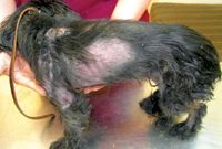
1. Chloe, a dog with pruritus, was referred for dermatologic evaluation after corticosteroids failed to resolve her pruritus. Yeast were identified on skin cytology, and treatment for Malassezia species was administered for 30 days.
3. You will not see as many organisms on a skin cytology sample as you would on an ear cytology sample. Finding one Malassezia organism every two or three oil fields on a skin cytology slide or even one field that has more than one organism supports a diagnosis of Malassezia dermatitis. In one study, normal dogs only had five organisms every 2,700 oil immersion fields,4 so even finding a few yeast is diagnostic.
It is beyond the scope of this article to go into all the treatments for dogs with pruritus, but a good take-home message is if you identify a dog with Malassezia dermatitis or a bacterial pyoderma, treat the infection before administering corticosteroids (Figures 1 & 2). Recheck the dog in 14 days to help you identify how much of the pruritus was due to secondary infection and how much (the remaining amount) is due to the primary disease.

2. Chloe 60 days after treatment for atopy (allergen-specific immunotherapy) and secondary Malassezia dermatitis (oral ketoconazole given once a day with food for 30 days; bathed with a 4% chlorhexidine shampoo followed by a humectant). No corticosteroids or other anti-inflammatories were given.
Paul Bloom, DVM, DACVD, DABVP (canine and feline)
Allergy, Skin and Ear Clinic for Pets
31205 Five Mile Road
Livonia, MI 48154
REFERENCES
1. Top 10 reasons pets visit vets. VPI Pet Insurance. Available at: http://www.petinsurance.com/healthzone/pet-articles/pet-health/Top-10-Reasons-Pets-Visit-Vets.aspx.
2. Outerbridge CA. Mycologic disorders of the skin. Clin Tech Small Anim Pract 2006;21(3):128-134.
3. Chen TA, Hill PB. The biology of Malassezia organisms and their ability to induce immune responses and skin disease. Vet Dermatol 2005;16(1):4-26.
4. Kennis RA, Rosser EJ Jr., Olivier NB, et al. Quantity and distribution of Malassezia organisms on the skin of clinically normal dogs. J Am Vet Med Assoc 1996;208(7):1048-1051.
Newsletter
From exam room tips to practice management insights, get trusted veterinary news delivered straight to your inbox—subscribe to dvm360.