CVC Highlights: Simple steps to proper periodontal probing
Don't forget this important procedure when performing a thorough oral examination.
Next >
Periodontal probing should be a part of every in-depth oral examination that is followed by professional dental cleaning. Measuring the depth of the gingival sulcus will help you determine the extent of a tooth's attachment, which will aid in deciding whether to extract a tooth, especially if dental radiography is unavailable to you.
Select the right probe
Various periodontal probes are available. I prefer using the CP-15 UNC probe (with millimeter markings and a wide, black marking at 5, 10, and 15 mm) in dogs and the Williams probe (with markings at 1, 2, 3, then 5, then 7, 8, 9, and 10 mm) in cats (Figure 1).
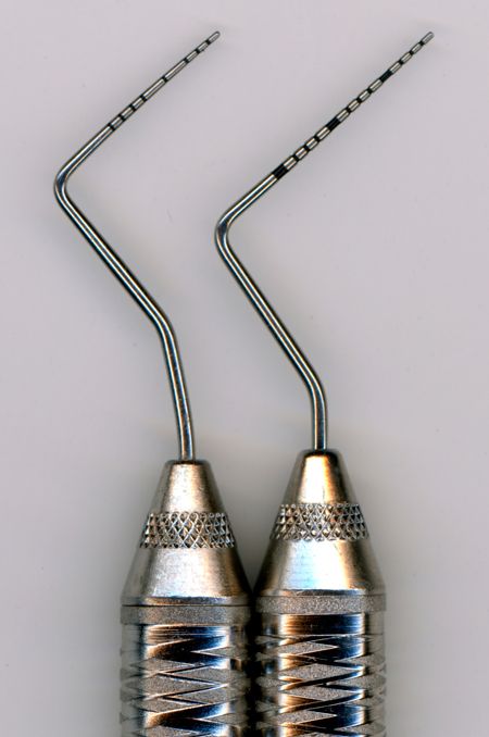
Figure 1: The Williams probe on the left (with markings at 1, 2, 3, then 5, then 7, 8, 9, and 10 mm) is preferred over the CP-15 UNC on the right (with millimeter markings and a wide, black marking at 5, 10, and 15 mm) for probing of feline teeth.
Get a grasp
To properly hold the probe, use a modified pen grasp. Put the instrument between your thumb and index finger (Figure 2A). Then bring your middle finger toward the probe's shank (Figure 2B).
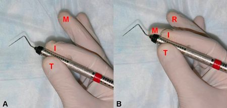
Figure 2A & 2B: The modified pen grasp; the instrument is first grasped between thumb and index finger (A), and then the middle finger is brought toward the probe's shank (B).
Support your hand with your ring finger while working inside the patient's mouth. By supporting your hand with your ring finger, you avoid slipping and possibly injuring the patient (Figure 3).
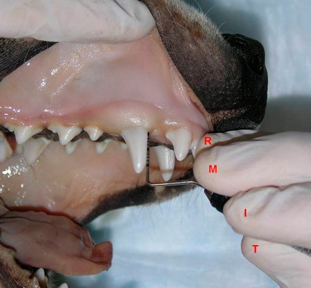
Figure 3: The modified pen grasp is used when probing the right maxillary canine tooth; the hand is supported with the ring finger on an incisor tooth.
Do not harm when probing the sulcus
Measure the gingival sulcus at a minimum of six points around each tooth's circumference-at three spots on the labial or buccal side and at three points on the lingual or palatal side. Do not keep the tip of the probe inside the sulcus when probing all around the tooth; doing so can seriously damage the gingival attachment.
Instead, walk the probe inside the sulcus. By applying gentle pressure, insert the probe, then withdraw it, insert it again a couple of millimeters farther away, withdraw it, and so on until you've probed around the tooth's circumference (Figure 4).
In dogs, the normal depth of the gingival sulcus is 1 to 3 mm. In cats, the normal depth is up to 0.5 mm.
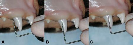
Figure 4: The periodontal probe has been inserted at the distolabial aspect of the right maxillary canine tooth (A); it is removed (B) and reinserted at the labial aspect of the tooth (C).
< Back
Don't stop with pocket depth
Remember, there's a difference between the pocket depth and attachment loss. You must also take gingival recession into consideration. For example, imagine an incisor tooth with a pocket depth of 6 mm and gingival recession of 2 mm. In this case, the total attachment loss would be 8 mm, and such a tooth would likely require extraction (Figure 5).
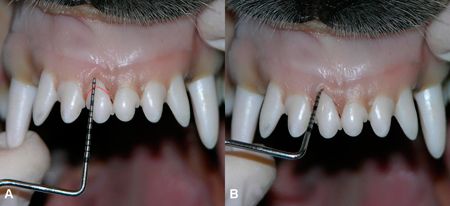
Figure 5: The difference between periodontal pocket and attachment loss is demonstrated in a dog cadaver; there is about 2 mm of gingival recession (A; dotted line indicating original gingival margin) and an about 6-mm pocket depth (B), totaling an 8-mm attachment loss.
Alexander M. Reiter, Dipl Tzt, Dr med vet, DAVDC, DEVDC
Dentistry and Oral Surgery Service Section of Surgery
Department of Clinical Studies
School of Veterinary Medicine
University of Pennsylvania
Philadelphia, PA 19104-6010