Dental corner: A goldendoodle with masticatory myositis
A veterinary team worked together to correct an autoimmune disorder that limits this dog's ability to open its mouth.
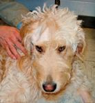
A 2-year-old neutered male goldendoodle named Gordon was presented to the University of Pennsylvania's Dentistry and Oral Surgery Service because he couldn't open his mouth. The owner reported that Gordon had been with a pet sitter while she was out of town and experienced no known trauma. When she noticed that Gordon was eating in a strange, messy way and that he couldn't open his mouth, she brought him to the emergency room.
Upon examination, Gordon was mildly dehydrated and drooling excessively, and his left temporomandibular area was enlarged. Gordon did not appear painful, but left-sided swelling and temporalis muscle wasting could be palpated. The owner could not tell whether Gordon had lost weight.
PHOTOS COURTESY OF THE UNIVERSITY OF PENNSYLVANIA
A complete blood count (CBC) and serum chemistry profile, including creatine kinase activity, were performed, and the results were normal. An intravenous catheter was placed, and anesthesia was induced with propofol. An oral exam revealed that Gordon's jaws couldn't be opened more than 1 inch. Since an endotracheal tube could not be placed by normal means, an emergency tracheostomy was performed. Gordon was taken for a computed tomography scan and then to the dental surgical suite for a more extensive oral exam.
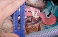
Photo 1: When Gordon was first examined, he could not open his mouth more than 1 inch.
A dental technician must become educated about diseases, such as masticatory myositis, and understand the anatomy.
Technicians assist in patient care preoperatively and postoperatively.
The extraoral exam
An extraoral exam revealed that the masticatory muscles were atrophied, and the doctors suspected chronic masticatory myositis because of the atrophy. This autoimmune disorder causes autoantibodies to attack certain muscle fibers (type 2M) found within the muscles of mastication—the temporalis, masseter, and pterygoid. It usually affects middle-aged and older large-breed dogs and results in atrophy of—and scar tissue deposition in—the affected muscles, leading to decreased range of motion of the jaws.
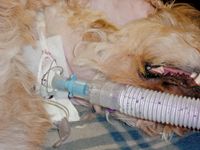
Photo 2: The anesthetist was unable to pass an endotracheal tube, even with a large laryngoscope, so a tracheostomy was performed.
Technicians must know how to process the required lab work and biopsy samples. In this case, three different biopsy samples were obtained, all requiring different containers and different processing fluids.
Treatment
Treatment involves high-dose corticosteroid therapy. A serum titer test was performed, and biopsy samples were obtained on the right side of the temporalis muscle because it was less inflamed.
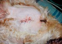
Photo 3: The doctor chose to perform a biopsy on the right temporalis muscle because it was less inflamed than the left side.
Gordon was given intravenous injections of cephalexin for the tracheostomy and biopsy and famotidine to reduce stomach acid. He was carefully monitored postoperatively, where he continued doing well; his tracheostomy tube was removed. Sucralfate (to be given before meals) to protect the stomach from the effects of high-dose corticosteroid therapy, tramadol, and immunosuppressive doses of prednisone (2 mg/kg) were prescribed. Gordon was able to lick water and baby food, and he was sent home with a high-protein soft diet.
A dental technician is also responsible for keeping detailed medical records for each patient.
Rechecks and follow-up care
At his one-week recheck, Gordon was eating and drinking well and able to open his mouth about 2 ½ inches. The biopsy and tracheostomy sites were healing normally. The prednisone dose was decreased to 1 mg/kg once a day. The 2M antibody test results were 1:4,000 (> 1:100 being positive). The muscle biopsy results were inflammatory myopathy/myositis with antibodies against type 2M fibers. The examination, computed tomography scan results, and treatment response alluded to the diagnosis, and the biopsy and antibody titer results confirmed it.
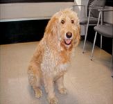
Photo 4: At his two-week recheck, he could open his mouth 4 ½ inches.
At a two-week recheck, Gordon could open his mouth 4 ½ inches but was demonstrating side effects of corticosteroid usage, such as polyuria, polyphagia, weight gain, and soft stools, so his prednisone dose was decreased to 0.5 mg/kg once a day. The famotidine and sucralfate were continued.
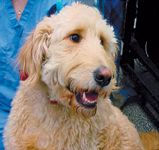
Photo 5: At his three-month recheck, Gordon smiles for the camera.
At a two-month recheck, Gordon was able to open his mouth enough to eat and drink normally and was able to hold small toys in his mouth. He had previously gained weight from corticosteriod use, and he was also back to his normal weight. His dose of prednisone was 2.5 mg once a day. He was encouraged to chew and play with toys to help strengthen his muscles. Six months later, the owner reported the dog was doing well.
Patricia March, CVT, VTS (Dentistry), is a dental technician at Animal Dental Center in Baltimore, Md., and the past president of the Academy of Veterinary Dental Technicians. March will teach on dental topics on the technician program May 11 at CVC Washington, D.C.Learn more and find wet lab opportunities at thecvc.com.