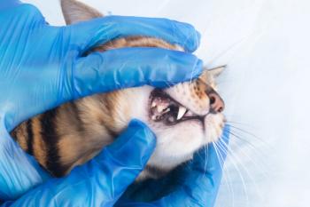
Dental Corner: Using intraoral regional anesthetic nerve blocks
Local anesthesia and regional anesthetic nerve blocks have been used for decades in human dentistry, but incorporating intraoral regional anesthetic blocks into veterinary dental and oral surgical procedures did not gain acceptance until the mid-1990s.
Local anesthesia and regional anesthetic nerve blocks have been used for decades in human dentistry, but incorporating intraoral regional anesthetic blocks into veterinary dental and oral surgical procedures did not gain acceptance until the mid-1990s.1-3 Regional anesthetic blocks, when combined with general anesthesia, provide preemptive analgesia to patients during and after painful procedures. Because regional anesthetic blocks produce complete analgesia before oral surgery, the amount of a general anesthetic needed to maintain a surgical plane of anesthesia is reduced. This reduction ameliorates the hypotensive and other adverse effects of inhalant anesthetics, and the savings from the reduced use of these inhalants (especially sevoflurane) over the course of a year can be great. Moreover, recovery from anesthesia is smoother when a patient has been provided complete analgesia, which reduces the chances of self or iatrogenic injury. And the need for immediate postoperative narcotics is also reduced or eliminated.
How regional anesthesia works
Regional anesthesia refers to blocking the nerve supply to a part of the body, as opposed to local anesthesia in which the anesthetic infiltrates the affected area. Intraoral regional anesthetic nerve blocks are used for a specific area of the mouth and can be used even if only one tooth in the area is to be operated on. Local anesthetic blocks, or splash or ligament blocks, may be used if only one tooth is to be operated on, but they are not as effective as regional blocks, in my experience.
The key to successful regional anesthesia lies in a thorough understanding of the appropriate neuroanatomy. Branches of the maxillary nerve supply sensory innervation to the maxillary teeth, and branches of the mandibular nerve innervate the mandibular teeth. The various regional blocks described below can be used alone or in combination to block sensory impulses from any or all areas of the dental arches.
Local anesthetics act on membrane channels to prevent nerve cell depolarization and to retard or prevent nerve impulse conduction. This mode of action is different from the analgesic properties of narcotics or nonsteroidal anti-inflammatory drugs, so the combination of local and systemic pain relief is not only complementary but also synergistic. The various local anesthetics differ in time of onset, duration of effect, and potential for toxicosis. An individual drug's effects can differ among species; for instance, cats generally have a lower threshold for toxicosis than dogs do. The local anesthetic of choice for intraoral regional anesthetic blocks is bupivacaine hydrochloride because of its prolonged duration of action. Although bupivacaine has a six- to 12-minute lag until the onset of analgesia, the analgesia will last four to six hours.
Figure 1. The proper needle placement for the cranial infraorbital nerve block. The local anesthetic is injected into the opening of the infraorbital foramen. Note the location of the foramen, apical to the distal root of the third premolar.
Performing the blocks
A small-gauge (25- or 27-ga), 1.5-in beveled hypodermic needle is required to minimize the trauma to nerves and blood vessels in the area of the block. Be sure to use a new needle, not the same one used to withdraw the anesthetic agent from the stock bottle. It is essential that aspiration for blood be done before any local anesthetic is injected. The potential for cardiac or acute central nervous system toxicosis greatly increases when these drugs are inadvertently administered intravenously. The total maximum dose of bupivacaine in a cat is 2 mg/kg, so do not inject more than 1 ml bupivacaine (0.5%) into an 11-lb (5-kg) cat during a single procedure. The maximum dose in dogs is 5 mg/kg. The toxic effects that can be observed in a patient that has received too much bupivacaine are cardiovascular arrhythmias and, less commonly, central nervous system signs.
Five regional anesthetic blocks can be used depending on the area of the oral cavity to be anesthetized. Except for the caudal mandibular block, the blocks are easy to learn. It is helpful to have a skull to refer to when performing a block to help visualize the anatomic landmarks. Make sure to insert the needle with the bevel facing out, and try to avoid repeatedly inserting the needle. Because of the lag with the onset of anesthesia, perform these blocks 10 to 15 minutes before beginning the procedure. The time between regional anesthetic injection and surgery can be used for scaling, oral and dental charting, taking intraoral radiographs, or performing other painless procedures. ä
Figure 2. As seen with this cat's skull, the position of the needle with the caudal infraorbital nerve block could cause orbital injury in a cat. For this reason, this block is not recommended in cats.
Cranial infraorbital nerve block
A cranial infraorbital nerve block anesthetizes only the ipsilateral incisors and canines of the maxilla. For this technique, 0.2 to 0.4 ml bupivacaine (0.5%) is injected into the opening of the infraorbital foramen, which is apical to the distal root of the maxillary third premolar. Palpate the foramen through the oral mucosa, hold the syringe almost parallel to the maxilla with the needle directed caudally, insert the needle 1 mm into the foramen, aspirate to ensure the needle is not in a blood vessel, and inject the bupivacaine rapidly (Figure 1). In cats, do not attempt to advance the needle into the foramen because the feline infraorbital canal is short, and needles placed into the foramen can cause orbital trauma.
Caudal infraorbital nerve block
A caudal infraorbital nerve block anesthetizes all ipsilateral teeth of the maxilla. This block is not recommended in cats because of the short canal and the potential for orbital injury (Figure 2). Insert the needle 2 to 3 mm into the infraorbital foramen, and place a finger over the entrance to the foramen. Inject 0.4 to 0.8 ml (usually 0.4 for small dogs, 0.6 ml for medium-sized dogs, and 0.8 ml for large dogs) bupivacaine (0.5%) into the foramen, causing the bupivacaine to flood the infraorbital canal and exit through the caudal opening of the canal (Figure 3).
Figure 3. The proper needle placement for the caudal infraorbital nerve block, which will block the entire unilateral maxillary arch on the side injected.
Middle mental nerve block
A middle mental nerve block anesthetizes all ipsilateral incisors and canines of the mandible. Inject 0.2 to 0.4 ml bupivacaine (0.5%) at the middle mental foramen, which is apical to the mesial root of the mandibular second premolar (Figure 4).
Figure 4. The proper needle placement for anesthetic injection at the middle mental foramen, which is apical to the mesial root of the mandibular second premolar.
Caudal mandibular nerve block
A caudal mandibular nerve block anesthetizes all ipsilateral teeth of the mandible. This procedure is challenging to perform accurately, so laboratory instruction is recommended before performing this block on patients.
Inject 0.8 to 1 ml bupivacaine (0.5%) at or near the mandibular foramen, which is on the lingual (inside) aspect of the mandible above the mandibular notch. The mandibular notch can be palpated caudal to the last molar on the ventral surface of the mandible. Insert the anesthetic needle through the skin, walk it off the ventral mandible, and advance it dorsally to the region of the mandibular foramen (Figure 5).
Figure 5. The proper needle placement for the caudal mandibular nerve block.
Palatine nerve block
The palatine nerve block partially anesthetizes the maxillary incisors, canines, and premolars. This block is recommended in cats. Keep in mind that most of the innervation to the maxillary arch comes from the infraorbital nerve, and the palatine block only offers some degree of anesthesia. Thus in dogs, the palatine nerve block should be performed simultaneously with the infraorbital nerve block to offer the highest degree of analgesia. Inject 0.1 to 0.2 ml bupivacaine (0.5%) at a midpoint between the mesial aspect of the maxillary carnassial tooth and the palatal midline (Figure 6).
Figure 6. The palatine nerve block. Local anesthetic is injected at a midpoint between the mesial aspect of the maxillary carnassial tooth and the palatal midline (arrows). One or both sides may be blocked simultaneously.
REFERENCES
1.
Anthony, J.: Intraoral regional anesthetic nerve blocks. Proc. World Veterinary Dental Congress, World Veterinary Dental Congress, Vancouver, British Columbia, 1995; pp 56-57.
2. Lantz, G.C.: Regional anesthesia for dentistry and oral surgery. J. Vet. Dent. 20 (3):181-186; 2003.
3. Holmstrom, S.E. et al.: Regional and local anesthesia. Veterinary Dental Techniques, 3rd Ed. (S.E. Holmstrom et al., eds.). W.B. Saunders, Philadelphia, Pa., 2004; pp 625-636.
"Dental Corner" was contributed by Daniel T. Carmichael, DVM, DAVDC, The Center For Specialized Veterinary Care, 609-5 Cantiague Rock Road, Westbury, NY 11590.
Newsletter
From exam room tips to practice management insights, get trusted veterinary news delivered straight to your inbox—subscribe to dvm360.


