
News|Articles|October 18, 2024
Diagnosing and treating canine struvite uroliths
Author(s)Austin Littrell, Assistant Editor

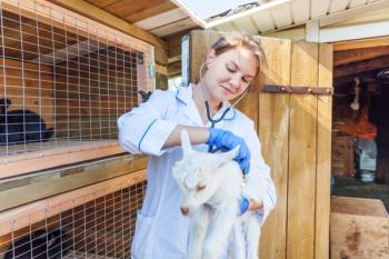
Common canine uroliths, including struvite, were covered during a session at the Fetch dvm360 Conference in Atlantic City
Advertisement
In his session, “Canine Uroliths,” presented at the Fetch dvm360 Conference in Atlantic City, New Jersey, Mark Acierno, MBA, DVM, DACVIM, professor and associate dean for clinical affairs at Midwestern University’s veterinary teaching hospital, detailed common canine uroliths, including methods for diagnosis and treatment. One of the uroliths covered was struvite, which is the most common urolith in dogs.
Generally, clinical signs of uroliths include lower urinary tract inflammation, hematuria, pollakiuria, periuria, and stranguria. In male dogs, the presentation of urethral uroliths can vary. Dogs may strain to urinate, but be unable to, and dribbling urine/intermittent incontinence could be observed.
Acierno advises attendees to palpate the bladder in every dog, and although the process will often be unrewarding, an audible crunching noise can occasionally be heard upon palpation, which indicates the presence of a urolith. He also recommends imaging the entire urinary tract, with radiology/ultrasound, also explaining that, in veterinary urology, “ultrasounds are always more important than getting x-rays.” According to Acierno, radiographs will show up to 27% false negatives, largely because of the size and composition of stones.
“Even in the hands of a boarded radiologist, 27% of the time, you are going to miss uroliths if all you’re looking at is x-rays,” Acierno said in his session. “[Using] ultrasound, you’re going to miss about 6%. So, ultrasound seems to be a better way to go.”
Advertisement
The best method for confirming the type of urolith is by collecting a piece of it and sending it off for laboratory analysis. For the removal, Acierno recommends several techniques, including urohydropropulsion, catheter-assisted retrieval, utilizing a stone basket, electrohydraulic lithotripsy, or laser lithotripsy.
Struvite uroliths
Struvite is the most common urolith in dogs. Composed of magnesium, ammonium, and phosphate, the stones are associated with urease-producing bacteria, including Staphylococcus intermedius, Proteus mirabilis, Pseudomonas spp., and Klebsiella spp. In dogs, struvite is almost always associated with urinary tract infections (UTIs). Meanwhile, in cats with struvite, stones are almost always sterile.
According to Acierno, urea is in the urine, and urease causes it to split into ammonium and bicarbonate. Ammonium then combines with magnesium and phosphate, and bicarbonate raises the pH of the urine. The increased pH results in decreased solubility of the ammonium, magnesium, phosphate complex, which causes struvite crystals and stones to form. Although females are anatomically more prone to urease-producing infections, struvite uroliths can become lodged in the long, narrow urethras of male dogs.
Treatment
A combination of antimicrobial and dietary therapy is required for the dissolution of struvite uroliths. Acierno recommends a discussion with the pet owner about pros and cons of medical and surgical management prior to any decisions regarding therapies. Antimicrobial therapy is based on a urine culture and sensitivity. Urine should be acquired through cystocentesis.
Acierno explains that bacteria continue producing urate, sequestered in the layers of the struvite stone, implying the importance of continued antibiotic therapy throughout the dissolution process. Struvite dissolving diets are low in protein, phosphate, and magnesium, and work to acidify the urine. He notes, though, that these diets are oftentimes not balanced nutritionally, and are not intended for long-term use.
Acierno sends pet owners home with pH test strips or urine dip-sticks so they can continue to monitor the pH level. “If the pH starts going back up, it means our [UTI] has either changed, or the bacteria has become resistant, and we’ve got a problem again,” Acierno explained. “I usually have them check a couple of times [per] week… if the pH is greater than 7, that suggests we have an issue and we need to get a urine sample [obtained by cystocentesis] submitted for culture.” He notes that complete dissolutions of struvite stones can take more than 3 months, and additional abdominal radiographs and urine analysis should occur monthly.
Prevention of recurrence
The prevention of struvite stones requires the prevention of alkaline urine, and therefore, a prevention of UTIs. Again, Acierno advocates for regular at-home urine pH measurements with either dip-sticks or a pH meter. He explained that the presence of alkaline urine would indicate that an office visit is needed, at which point urine should be collected by cystocentesis, and submitted for culture. If necessary, he also recommends radiographs during this visit. For recurrent UTIs, Acierno advises evaluating for structural abnormalities that would predispose the patient to UTIs. Oftentimes, these cases are related to involuted vulvas, which can be corrected surgically. Other patients with recurrent UTIs could benefit from long term, low-dose antibiotic therapy.
Reference
Acierno M. Canine Uroliths. Presented at: Fetch dvm360 Conference; October 14-16, 2024; Atlantic City, NJ.
Newsletter
From exam room tips to practice management insights, get trusted veterinary news delivered straight to your inbox—subscribe to dvm360.
Advertisement
Related Content
Advertisement

Advertisement
Advertisement