Diagnostic Imaging: The subtleties in identifying a bronchial pattern
A bronchial pattern on radiographs indicates pathology involving the airways. It can be a subtle pattern to recognize, so let's look at some of the features.
A bronchial pattern on radiographs indicates pathology involving the airways. It can be a subtle pattern to recognize, so let's look at some of the features.
Normal bronchi
The airways are made of cartilage that is radiolucent, but they have some surrounding soft-tissue structures that can make them visible. The bronchi are normally seen near the heart base, where the diameter is still quite large.
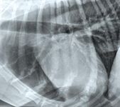
You might see parallel thin walls with vessels on either side, or just see the airways flanked by the pulmonary arteries and veins (Image 1). The bronchi are visible near the hilus, but the walls are thin and sharp and are not visible toward the periphery.

Image 1: Airways flanked by the pulmonary arteries and veins.
Mineralized bronchi
With age, many dogs develop mineralization of the airways and trachea. It is not a normal aging change in cats. Mineralized bronchi are recognizable because of their mineral opacity and very thin, sharp borders. The bronchi are visible farther out in the periphery than in a dog with no mineralization. But any increase in opacity is uniform and very opaque.
Bronchial pattern
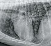
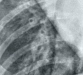
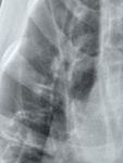
In a true bronchial pattern that stems from infectious/inflammatory disease, the bronchial walls are thickened because of inflammatory tissue and cells surrounding the airways. This makes them easier to see, especially in the periphery of the lung (Image 2). The parallel lines you see are called tram tracks, and a bronchus visible end-on with thickened walls is called a donut. It also can be described as a cygnet ring, because it tends to be of non-uniform thickness (Image 3). Additionally, the bronchial walls are not as sharp or regular as normal or mineralized bronchi (Image 4). Chronic bronchial disease can result in bronchiectasis, which is an irreversible dilation of the bronchi. They don't taper toward the periphery and can be saccular in appearance.

Image 2: Bronchial walls thickened by inflammation are easier to see.
Causes of bronchial patterns
Differential diagnoses for a bronchial pattern usually are inflammatory (chronic bronchitis, eosinophilic infiltrates, parasitic infections, fungal or bacterial pneumonia). Pulmonary edema sometimes makes the bronchi look thickened, because the tissues surrounding the airways fill with fluid. Identifying additional abnormalities can help you rank your differentials appropriately. For example, a bronchial pattern and large perihilar lymph nodes would point toward fungal pneumonia.

Image 3: A bronchus viewed end-on with thickened walls is called a donut.
Identifying abnormal bronchi
Once you have ruled out normal large airways and benign bronchial mineralization as the cause for increased visibility of bronchi, search for the hallmarks of the pattern. Bronchial thickening can be a subtle finding, especially in cats. Make sure to look in the periphery for donuts, especially on the v/d or d/v projection where they show up better. Tram tracks often are best seen overlying the diaphragm or heart, where the summation effect makes them more apparent (Image 2). A magnifying glass can be helpful to search for small airways.

Image 4: Thickened bronchial walls are indistinct and visible in the periphery of the lung.
Dr. Zwingenberger is a veterinary radiologist at the University of California-Davis.
Veterinary scene down under: Australia welcomes first mobile CT scanner, and more news
June 25th 2024Updates on the launch of the first mobile CT scanner available for Australian pets; and learn about the innovative device which simplifies placement of urinary catheters in female dogs
Read More