Don't be fooled by look-alike skin diseases
All of us, at one time or another, were probably guilty of treating one of our sarcoptic mange patients as an allergic patient. It is the perfect example of a patient with the same clinical appearance and symptoms of two diseases: atopy vs. sarcoptic mange.
All of us, at one time or another, were probably guilty of treating one of our sarcoptic mange patients as an allergic patient. It is the perfect example of a patient with the same clinical appearance and symptoms of two diseases: atopy vs. sarcoptic mange. The difficulty in treating most dermatological cases is, in fact, that the same disease can appear differently in different patients. Conversely, two patients that appear to have the same clinical appearance may, in fact, have two different diseases. There are many examples of this dilemma which we deal with daily, but probably the most serious look-alike diseases are bacterial pyoderma vs. cutaneous lymphoma and pemphigus foliaceus vs. bacterial or dermatophyte infections. (See Photos 1 and 3.)
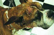
Photo 1: Cutaneous lymphoma in a Boxer resembling a spreading bacterial pyoderma.
Cutaneous lymphoma
Understandably, mistaking a bacterial pyoderma for cutaneous lymphoma results in serious consequences due to the extremely differing prognoses of the two diseases.
Cutaneous lymphoma is usually a disease of older dogs and cats with the most common breeds seen in our practice being Boxers and Golden Retrievers (see Photo 2). Cutaneous lymphoma can be epitheliotropic (usually T lymphocyte in origin in the dog) or non-epitheliotropic (usually B lymphocyte in origin). Sometimes referred to as mycosis fungoides, cutaneous lymphoma can have four presentations-exfoliative (see Photo 4, p. 5S) (the patient presents with erythema and a "peeling" of the skin sometimes resulting in large flakes), plaque (see Photo 5, p. 5S) (presents as nonraised, sometimes spherical shaped inflamed lesions), tumor stage (see Photos 6, 7, p. 5S) (usually varying sizes of erythemic raised, almost plateau-like lesions) and mucocutaneous ulceration/erythema.
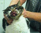
Photo 2: Cutaneous lymphoma in a feline.
Resembling bacterial pyoderma
The first three of these presentations may resemble a bacterial pyoderma and in fact, a bacterial pyoderma may accompany these cutaneous lymphoma lesions. The patients have varying degrees of pruritus, are afebrile, with or without lymphadenopathy.

Photo 3: Bacterial pyoderma, dermatophytosis or pemphigus foliaceus in a Dachshund.
Perhaps the key "clue" is the nonresponsiveness to antibiotics and the spreading of lesions. An in-office cytology can be helpful in determining the next diagnostic step-if lymphocytes are seen on cytology or if the patient is not responding to antibiotics, skin biopsies should be performed. As mentioned, secondary pyodermas may accompany cutaneous lymphoma lesions and neutrophils along with lymphocytes may be seen on cytology.
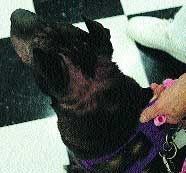
Photo 4: Canine with generalized erythema of skin due to epitheliotopic lymphoma. Note the "raspberry" color of the skin often seen in cutaneous lymphoma.
Be sure to obtain several skin biopsies of different stages of the lesions to achieve an accurate diagnosis and include a thorough history and response to therapy for the pathologist. If, for any reason, you do not feel the skin biopsy results match the patient's clinical appearance, rebiopsy, as representative samples may not have been submitted.
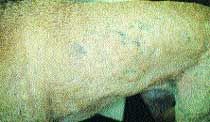
Photo 5: Plaque stage of cutaneous lymphoma resembling a bacterial pyoderma.
Unfortunately the prognosis for cutaneous lymphoma is grave despite medications such as prednisone, retinoids, lomustine, vincristine, adriamycin, and cytoxan. However making the correct diagnosis as soon as possible may buy the patient a bit more time and save the owner unnecessary expenses for medications that were not indicated.

Photo 6: Focal tumor stage of cutaneous lymphoma.
Separating diseases
In the dog and cat, differentiating between pemphigus foliaceus (PF) (see Photos 8, 9, p. 19S) and a bacterial or dermatophyte infection can be difficult, both clinically and sometimes histologically. Both diseases can affect any age and breed. Pemphigus patients may be febrile with nondescript laboratory parameters such as neutrophilia and hyperglobulinemia. Clinically, multi-focal crusting or pustules may occur on the ear pinna, face, trunk, nipples or footpads.

Photo 7: Multifocal tumors of cutaneous lymphoma.
In the dog with a bacterial pyoderma, cytology of an intact pustule may yield degenerative neutrophils with or without bacteria and few, if any, acantholytic cells.
Culture and sensitivity yields normal skin flora such as staphylococcus intermedius. In a patient with PF, cytology of an intact pustule may yield degenerative neutrophils, eosinophils, acantholytic cells and an absence of bacterial organisms. Culture and sensitivity of a pustule yields no organisms.
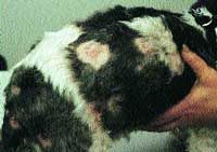
Photo 8: Multifocal pemphigus foliaceus lesions in a dog.
Skin biopsies are necessary to make the diagnosis of PF vs. bacterial pyoderma but because of the similarities, it may be difficult as well for the pathologist. (See Photo 10.)
Be sure to include a good history and response to therapy with the biopsy samples. Ideally, intact pustules if present, are good submissions, but dried pustules may yield acantholytic cells as well. Skin biopsies for culture may also be helpful as well as samples for immunohistochemistry. Fungal cultures and trichograms should always be performed when pemphigus foliaceus is a differential since Tricophyton spp. (see Photo 11) can result in acantholytic cells.
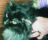
Photo 9: Crusty pinna in a cat with pemphigus foliaceus.
It is extremely important to differentiate between PF which is an immune-mediated disease and needs to be treated as such, and an infectious disease be it bacterial or fungal which is treated with the exact opposite medications! Do not start the patient on steroids before the skin biopsies are performed as in the case of PF, it may negate the histopathological changes necessary to be seen in the biopsy to confirm the disease. If steroids are used in a bacterial or fungal infection, the infection will only be made worse and the chance for bacterial septicemia occurs.
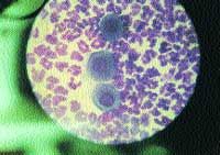
Photo 10: Large purple staining acantholytic cells seen in a cytology of a pustule in a patient with pemphigusfoliaceus. Acantholytic cells are "rounded up" epithelial cells.
Everyday, each patient and every disease is a challenge for every veterinarian. However perhaps dermatology can be the most frustrating due to the same disease presenting differently and vice versa. When faced with a dermatology patient whom you feel is not responding to therapy, do not hesitate to biopsy and consult your local veterinary dermatologist.

Photo 11: A trichogram showing fungal spores completely invading a hair shaft in a patient with dermatophytosis.