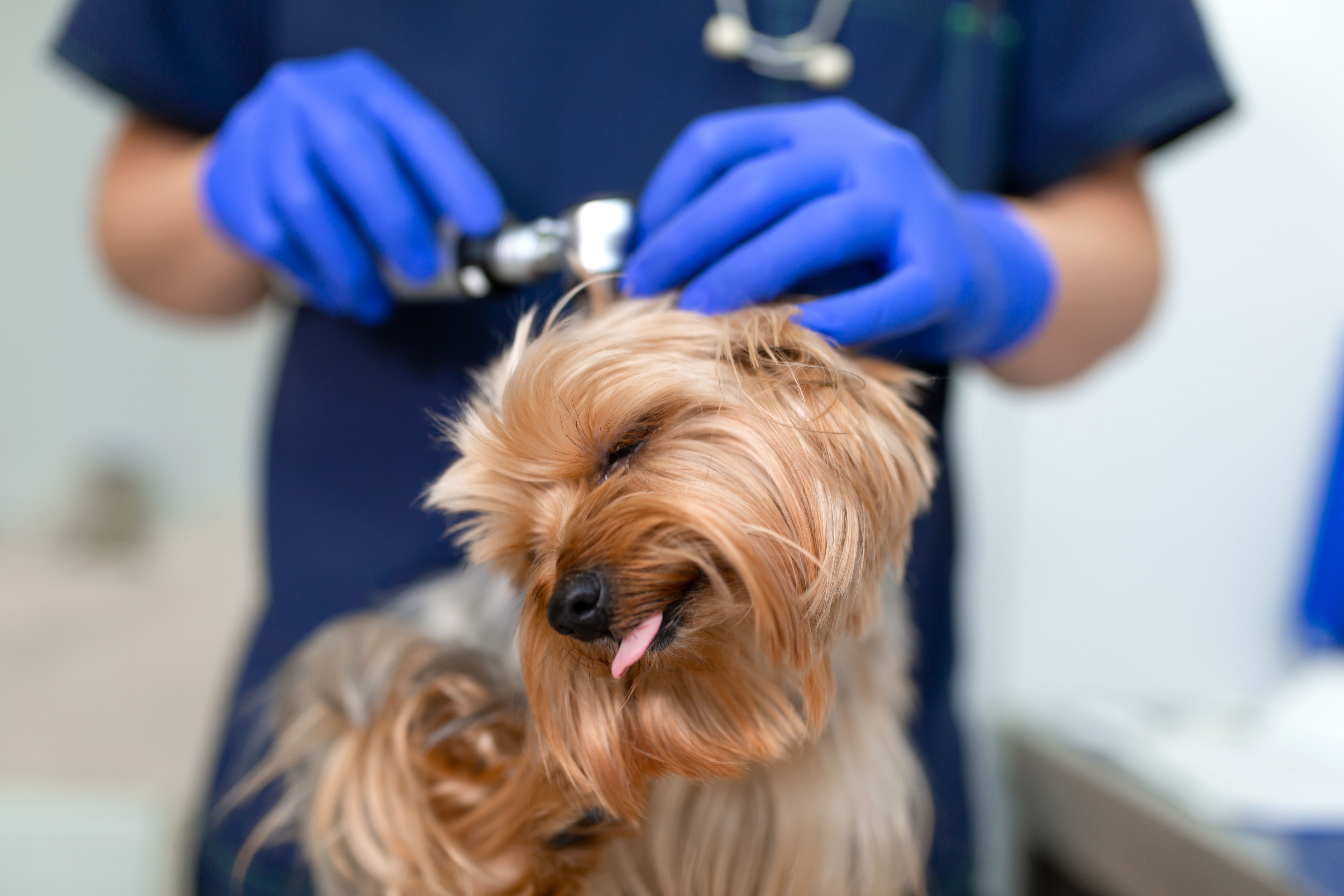
It’s been said that only two things are certain in this world: death and taxes. But if we’re talking about the veterinary world, that list should arguably expand to include otitis. “We know that otitis is the second most common reason dogs come into our clinics,” said Ashley Bourgeois, DVM, DACVD, at a recent Fetch dvm360 conference session. “The first reason is itch, so derm is awesome because we get the No. 1 and 2 spots! And usually it’s both—they have an ear infection because they have allergies and they’re itchy.”
But despite all of this forced practice, diagnosing otitis remains a painful process for both the pet and the practitioner—a reality that Dr. Bourgeois addressed by sharing her solutions to some common conundrums.
Problem: The patient is extremely uncomfortable during the otoscopic exam.
Treatment: The patient may just need a distraction, and you may need to tweak your technique.“ There actually aren’t too many dogs, unless they’re extremely painful or just aggressive on their own, that I can’t do an awake otoscopy exam on,” said Dr. Bourgeois. “Obviously I’m doing them all day, every day, so I have good practice, but we’ll use things to distract them.” If the dog doesn’t have a food allergy, her diversion weapon of choice is a pretzel rod garnished with spray cheese.
But distraction is only one part of a better otoscopy exam. “Dogs tend to get uncomfortable when you put in the scope and hit the junction where the vertical canal meets the horizontal,” explained Dr. Bourgeois. “Because the ear canal is L-shaped, you’ll want to pull it up and out to straighten the canal. You’ll be able to get deeper into the canal, and it will be less painful for the dog.”
One more tip on otoscopy: Keep more than one size of speculum in your clinic—preferably three. “Three-millimeter cones are the most common, but we have at least three different sizes in our clinic because you don’t want to use the same size cone on a Chihuahua as you do on a Great Dane,” said Dr. Bourgeois. “You want the biggest area of visualization you can get, but you’re not going to be able to jam the bigger cone into the Chihuahua’s ear.”
Problem: You suspect there’s an infection, but the culture came back negative.
Treatment: Perform a cytologic examination before you culture. Dr. Bourgeois didn’t mince her words: “Always do a cytology. Always. I don’t care if you’re seeing a recheck and you think it looks a lot better.” She explained that the exam provides a quick overview of the aural environment, as well as a foundation for therapeutic decisions and advanced diagnostics, and is the primary tool for identifying bacterial or yeast overgrowth. Accordingly, Dr. Bourgeois said that a cytologic exam should be performed before culture and sensitivity (C/S) testing because if only yeast is noted, bacterial C/S testing isn’t recommended. “Who wants to get a negative culture and then go back and perform a cytologic exam only to find it was just yeast to begin with? Culturing is not inexpensive, so you don’t want to waste the client’s money when it’s only yeast that’s present,” she advised.
Performing a cytologic exam can also prove useful if the results of your culture don’t end up matching what you saw. “Have you ever gotten tons of rods and you’re for sure going to grow Pseudomonas, only to get your culture back and have it say, ‘1+ Staphylococcus’?” asked Dr. Bourgeois. “You need to make sure you’re doing cytology and correlating it with the culture results because cultures can have issues too.” If the two don’t match up, you can call the lab to see if an error occurred.
If you’re unsure how to best approach evaluating a cytology slide, Dr. Bourgeois has advice to offer. “I always suggest scanning at a lower power first to look for representative areas on the slide,” she explained. “You want to look for areas that have inflammation present but that aren’t just masses of cells on top of themselves. Avoid the globs.” Once you find an area, Dr. Bourgeois continued, you can move into a higher power.
Problem: The patient’s ear is hidden behind layers of gunk and biofilm.
Solution: Consider a thorough cleaning as part of your workup. Being able to visualize the ear is a vital component of the diagnostic exam, which means you may need to spend some time removing debris so you can really see what’s going on. Perhaps, for example, the primary cause of a patient’s otitis is a polyp that’s hidden under several layers of gunk and biofilm. Flushing can also help you identify small tears in the tympanic membrane, said. Dr. Bourgeois. “If you see a bubble coming up through the tympanic membrane, that’s an indication you have a microtear.”
Dr. Bourgeois uses red rubber catheters and infant feeding tubes for deep ear flushes. “In the handheld scope,” she noted, “I will try to use an 8-French catheter. You can’t get anything that big in a fiberoptic video-enhanced otoscope, so I use a 5-French catheter.” 5-French catheters have gone on and off backorder for a while, so Dr. Bourgeois has had to use polypropylene instead. “But you have to be careful with polypropylene catheters because they’re more rigid and can indent and cause a myringotomy,” she warned. “In fact, that’s what we use them for.” And while she doesn’t typically use general anesthesia for otoscopic exams, she noted that it can be very helpful for deep ear flushes, when possible.
When asked how long to wait between flushing the ear and applying topical therapy, Dr. Bourgeois recommended a period of five to 10 minutes. “I like to give the flush time to remove debris and clear away so the drop can penetrate the canal better, but you could probably apply the topical right after and be fine.”
Culturing the middle ear
“If you’re concerned about a middle ear infection, you definitely need to culture the middle ear,” said Dr. Bourgeois. “And if you don’t feel comfortable with it, try to get them referred.” She typically uses a polypropylene catheter through her video otoscopy unit, though she noted that you can use a sterile plastic catheter or spinal needle if using a handheld scope. “If the eardrum is intact, we’ll do a myringotomy. We usually cut the end of the polypropylene catheter at an angle to make a little point and then we push it into the tympanic membrane until we get that lovely popping sound and it’s gone through,” Dr. Bourgeois explained. If the membrane is really stiff, it can take some time. “Don’t keep trying to jam it in. Use slow and steady pressure.”
Sold on cytology
Dr. Bourgeois closed the session with a friendly reminder shared with the earnestness of someone who has learned a lesson the hard way: “If you take nothing else away today, please perform a cytologic exam every time.” She’s often shocked by what she finds in seemingly “normal” ears.
C/S testing is indicated when:
A mixed infection is present. “For example, if you see a lot of cocci and rods on cytology, you need to find the sensitivities for those because it’s not normal for a dog to have an abundance of bacteria and numerous types of bacteria,” she explained.
Suppurative inflammation (including with bacterial rods, cocci, or no visible organisms) is revealed during initial cytology. In such cases, you need to get to the bottom of what’s going on quickly, said Dr. Bourgeois.
Otitis media is suspected. “These are cases that need systemic therapy,” she noted. “Remember that dogs have L-shaped ear canals, so to rely on an owner to be able to put a drop in the canal, have it travel all the way down the vertical canal, all the way down the horizontal canal and then get to the bullae isn’t realistic.”
There’s no response to appropriate topical and systemic antibiotic therapy.
If the eardrum isn’t intact, she injects sterile saline into the middle ear and then aspirates it back up. “And that aspirate is what we culture,” continued Dr. Bourgeois. “We don’t culture the tube because it went through all of the external ear canal.”
