The effect of hydration status on echocardiographic measurements of normal cats
In a recent article (The Effect of Hydration Status on the Echocardiographic Measurements of Normal Cats, by Campbell, F.E. & Kittleson, M.D. J Vet Intern Med 2007), the authors examined the effect of hydration status on echocardio-graphic findings in normal cats.
In a recent article (The Effect of Hydration Status on the Echocardiographic Measurements of Normal Cats, by Campbell, F.E. & Kittleson, M.D. J Vet Intern Med 2007), the authors examined the effect of hydration status on echocardio-graphic findings in normal cats.
The study was designed to parallel commonly encountered clinical situations.
They found that hydration status can significantly alter two-dimensional echocardiographic measurements used in the diagnosis of most heart diseases in cats.
Table 1: Check parameters, then diagnose
These changes in echocardiographic parameters are enough to potentially confound or mask the presence or severity of any of the five main feline cardiomyopathies — hypertrophic cardiomyopathy, dilated cardiomyo-pathy, restrictive cardiomyopathy, unclassified cardiomyopathy and arrhythmogenic right ventricular cardio-myopathy.
"Normal" cats, therefore, potentially can be misdiagnosed as having heart disease, and those with heart disease may be incorrectly diagnosed as being free of heart disease, or the extent of their disease underestimated.
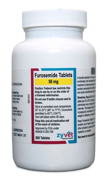
In this study, volume depletion was achieved through the administration of furosemide while temporarily delaying food and water intake.
Dehydration resulted in pseudo-hypertrophy, reduced left atrial size and left ventricular end-systolic chamber obliteration.
A state of increased circulating volume was achieved by administering intravenous fluids at an accepted anesthetic/surgical rate (10ml/kg/hr).
Increasing the circulating volume resulted in increased left ventricular and left atrial dimensions, and in some cases the development of a heart murmur.
These alterations in chamber size artifactually changed the left artrial/aortic root ratios, which could therefore result in a spurious clinical diagnosis.
Table 1 compares the left atrial:aortic root of normal cats (from Small Animal Diagnostics Ultrasound, Nyland/Mattoon, 2002) to those from the article.
This study emphasizes the importance of assessing a patient as a whole before performing or interpreting an echocardiogram.
The clinician should consider correcting the hydration status in cats (and probably dogs) prior to performing and interpreting an echocardiogram.
The clinician must realize that ultrasonographic imaging, as well as other advanced diagnostics, never stand alone.
They are just a part of the complete diagnostic effort.
Dr. Runde is a 2001 graduate of the University of Pennsylvania School of Veterinary Medicine. He completed an internship at Hollywood Animal Hospital. He is an associate veterinarian at the Animal Emergency and Referral Center, Ft. Pierce, Fla., where he has been an internal medicine resident since 2007.
Newsletter
From exam room tips to practice management insights, get trusted veterinary news delivered straight to your inbox—subscribe to dvm360.


