Feline eye implants offer hope for humans
Columbia, Mo. - A University of Missouri (MU) veterinary ophthalmologist hopes continued research and advancement in eye-implantation technology will one day end retinal blindness for companion animals and humans.
COLUMBIA, MO. — A University of Missouri (MU) veterinary ophthalmologist hopes continued research and advancement in eye-implantation technology will one day end retinal blindness for companion animals and humans.
"About one in 3,500 people worldwide is affected with the hereditary disease retinitis pigmentosa that causes the death of retinal cells and, eventually, blindness," says Kristina Narfstrom, DVM, PhD and MU College of Veterinary Medicine veterinary ophthalmologist.
Narfstrom partnered with Optobionics Corp. in Naperville, Ill., and Machelle Pardue, a researcher with Emory University and the Research Service at the Atlanta VA Medical Center to create a feline eye implantation to improve vision and protect retinal cells from the disease.
Having studied retinal failure and feline retinal surgery for more than 10 years, Narfstrom brought her veterinary expertise to the project in spring 2006.
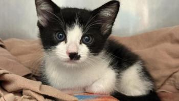
Working with Abyssinian and Persian cats, breeds affected by hereditary retinal blinding disease, Narfstrom has seen improved vision in all 11 implantations she has performed on severely visually impaired or blind cats. Feline eyes approximate those of humans in size and construction, allowing surgeons to use the same techniques and equipment on both.
"Our current study is aimed at determining safety issues in regard to the implants and to further develop surgical techniques. We also are examining the protection the implants might provide to the retinal cells that are dying due to disease progression, with the hope that natural sight can be maintained much longer than would be possible in an untreated patient," Narfstrom says.
During surgery, Narfstrom makes two small cuts into the sclera, the outer wall of the eyeball.
After removing the vitreous, which is the gelatinous fluid inside the back part of the eyeball, she creates a small blister in the retina and a small opening, large enough for the microchip. Just two millimeters in diameter and 23 micrometers thick, the chip includes several thousand microphotodiodes that react to light and produce small electrical impulses in parts of the retina.
The minimally invasive surgery takes less than two hours and has a fairly quick recovery time — only several days —Narfstrom says.
"In the future, I hope this will be one way of treating end-stage blindness in dogs and cats, but the timeline is difficult to say. It could be a couple of years before we know if this works well, so it is really still in the research phase. But I have hopes it is something we can practically do in the future," Narfstrom says.
Working off a MU grant and funding from Optobionics, Narfstrom says she is concerned about how continued progress and research will be supported in the future. But the project already has yielded positive results in humans.
"There are already clinical trials going on in humans. They started six years ago, and now 30 people have been operated on," Narfstrom says. "But they are trying to make this implant better all the time, and that is where I come in."
Newsletter
From exam room tips to practice management insights, get trusted veterinary news delivered straight to your inbox—subscribe to dvm360.


