
|Articles|February 1, 2012
Fine-needle biopsy: Are you using this valuable diagnostic tool?
A recent article discussed how fine-needle aspiration and fine-needle fenestration can be powerful diagnostic tools.
Advertisement
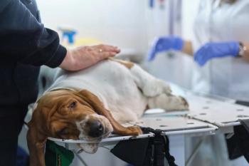
A recent article discussed how fine-needle aspiration (FNA) and fine-needle fenestration (FNF) can be powerful diagnostic tools.1 In short, FNAs and FNFs are most useful in obtaining diagnostic samples of neoplastic and infectious processes and less useful in chronic inflammatory processes (e.g., cholangiohepatitis, cirrhosis). In many situations, they can be used to make a definitive diagnosis with considerably less risk to the patient and at a considerably lower cost to the owners.
If a definitive diagnosis is not reached, FNA or FNF can often help refine the differentials. Full-thickness, or wedge, biopsy samples are often needed for a definitive diagnosis. Collecting representative cells is key, as poor-quality samples can be not only nondiagnostic but also misleading, resulting in inappropriate therapy or diagnostics.
Reducing the risk of complications
Complications involved with FNA or FNF of internal organs and bone are a valid concern and may discourage many clinicians. But, in fact, human and veterinary medicine studies have shown low complication rates. And measures can be taken to drastically reduce complication rates.
Risk of hemorrhage can be reduced by obtaining samples under ultrasound guidance and assessing hemostasis in situations in which the lesion of interest is highly vascular or when a coagulopathy is suspected. Seeding along the needle track of tumor cells is another common concern, but the risk of this occurring is quite small. The incidence of pneumothorax after transthoracic aspiration (requiring therapeutic intervention) is low and can be reduced by using ultrasound guidance and sedation.
In general, complications can be reduced by using correct technique, ultrasound guidance, appropriate sedation and pain management and sound clinical judgment on a case-by-case basis.
Efficacy and indications
Advertisement
FNA and FNF can be used with varying diagnostic yield in a variety of organs, particularly in bones, lungs and pleural fluid (Table 1).
Table 1: FNA and FHF utility in various organs
Bone cytology, according to the study, correlates with histopathologic diagnosis in about 70 percent of the cases, and in cases of bone tumors, the correlation is even higher (92 to 97 percent sensitivity and 100 percent specificity for sarcomas).1 Cytology is less helpful for nonneoplastic bone lesions. Alkaline phosphatase stain is useful to differentiate canine osteosarcoma from other sarcomas. This stain is highly sensitive and specific for osteosarcoma, with a sensitivity of 100 percent and specificity of 89 percent.
FNA or FNF of pulmonary lesions can be rewarding and diagnostic for pulmonary blastomycosis or neoplasia. Pulmonary lesions that are focal and peripherally located near the body wall are the easiest to evaluate (Figure 1).
Figure 1: This pulmonary lesion located near the body wall is an ideal candidate for FNA or FNF.
Pleural fluid is most often easily obtained and can be both therapeutic and diagnostic. Cytology can help differentiate among pyothorax, hemothorax, chylothorax and neoplastic processes.
In many cases, obtaining samples from intra-abdominal organs via FNA or FNF can result in a definitive diagnosis, so clinicians can often avoid more invasive and expensive testing. If the results are nondiagnostic, information obtained from these samples may help refine the differential list. Samples can also be used for culture and sensitivity if an infectious process is suspected.
A few tips
When submitting samples for review, verify that the sample is representative of the lesion. It is helpful to make several slides from multiple locations. If blood contamination is an issue, use a smaller gauge needle for sampling.2
Be sure to inform the owner verbally and in writing that in many cases (especially in cases of liver disease) the aspirated sample may not represent the causative disease process and may also be nondiagnostic. Providing unstained samples will allow cytologists to choose their stains of choice. An accurate history and other clinical information (i.e., images of the area in question) are also valuable to consulting pathologists.
Dr. Lyman is a graduate of The Ohio State University College of Veterinary Medicine. He completed a formal internship at the Animal Medical Center in New York City. Lyman is a co-author of chapters in the 2000 editions of Kirk's Current Veterinary Therapy XIII and Quick Reference to Veterinary Medicine.
Dr. Runde is a graduate of the University of Pennsylvania School of Veterinary Medicine. He completed an internship at Hollywood Animal Hospital. He is an associate veterinarian at the Animal Emergency and Referral Center in Ft. Pierce, Fla.
References
1. Wypij JM. Getting to the point: indications for fine-needle aspiration of internal organs and bone. Top Companion Anim Med 2011;26(2):77-85.
2. Rogers KS. Collection of specimens for cytology. In: Bonagura JD, Twedt DC, eds. Current veterinary therapy XIV. St. Louis, Mo: Saunders Elsevier, 2009;301-304.
Newsletter
From exam room tips to practice management insights, get trusted veterinary news delivered straight to your inbox—subscribe to dvm360.
Advertisement
Related Content
Advertisement
Advertisement
Advertisement
Trending on dvm360
1
Conference Insider: VMX will celebrate “champions of care” in 2026
2
Tarter control toothpaste receives VOHC Seal of Acceptance
3
Q&A: What the 2023 ACVIM update means for leptospirosis vaccination
4
The hidden cost of staying strong: How emotional armor protects you, and what it costs
5