Gastric neoplasia best treated surgically
Canine, Labrador Retriever/Collie cross, 7 years old, male neutered, 57 lbs.
Signalment:
Canine, Labrador Retriever/Collie cross, 7 years old, male neutered, 57 lbs.
Clinical history:
The dog presents with poor appetite and moderate lethargy for two months and weight loss. The results of the Ehrlichia canis and Lyme disease tests are negative and the Rocky Mountain spotted fever test is positive at 1:128. Therapy has included tetracycline.
Physical examination:
The findings include rectal temperature 102.6° F, heart rate 120/min, slightly pale mucous membranes, and normal capillary refill time. Normal heart and lung sounds are heard. Abdominal distension, thinness, and generalized muscle wasting are noted.
Laboratory results:
A complete blood count, serum chemistry profile and urinalysis are outlined in Table 1.
Ultrasound examination:
Thorough abdominal ultrasonography was performed.
My comments:
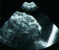
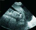
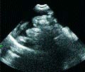
There is a moderate amount of free fluid accumulated within the abdominal cavity. The liver shows a uniform echogenicity in its parenchyma. No masses noted within the liver parenchyma. The gall bladder is mildly distended, and its walls are not thickened or hyperechoic. The spleen shows a uniform echogenicity in its parenchyma - no masses noted. The left and right kidneys are similar in size, shape and echotexture. No masses or calculi were noted in either kidney. The urinary bladder is distended with urine and contains some urine sediment material - no masses or calculi noted. The left and right adrenal glands are similar in size and shape. The stomach wall is irregularly thickened and echobright. The small intestinal wall is normal. The pancreas is enlarged.

Table 1: Results of laboratory tests
Cytologic examination:
About 1,300 milliliters of ascitic fluid was removed prior to further ultrasonographic evaluation. Fine needle aspirations of the stomach wall and sample of ascitic fluid for cytologic examination were collected and submitted for laboratory evaluation.
The cytologic results indicate gastric wall adenocarcinoma and neoplastic effusion secondary to disseminated carcinoma (carcinomatosis).
Case management:
In this case, gastric wall adenocarcinoma with metastatic spread is the clinical diagnosis. Clinically, there is not much one can do for this case with this histopathologic diagnosis. Chemotherapy may be attempted but carries a grave prognosis.
Review of gastric neoplasia
Gastric neoplasia accounts for less than 1 percent of all canine malignancies. Adenocarcinoma is the most common type, affecting older (average age between 7.5 to 10 years) male dogs.

Image 1.
Other malignant tumors include leiomyosarcoma and lymphoma. Benign tumors (adenomas and leiomyomas) are uncommon and affect older dogs (average age being 16 years).
Lymphosarcoma is the most common gastric neoplasm in cats. Gastric adenocarcinomas are most common on the lesser curvature and pylorus and often involve most of the gastric body. Metastases are common, especially in the gastric lymph nodes, but also in the spleen, liver, omentum, peritoneum and lungs. Gastric lymphosarcoma may appear as a single mass or multiple, often ulcerated nodules, or as a diffuse infiltrative lesion. Regional lymph nodes are often involved. Adenomas present as polypoid masses, which may occlude the pylorus. Leiomyomas present as single or multiple sessile, firm polyps, covered by a normal mucosa. They are localized in the gastric body or near the cardia.

Image 2.
Diagnostic considerations
Dogs with gastric adenocarcinoma usually present with vomiting, anorexia and weight loss. The duration of clinical signs is from weeks to months. Definitive diagnosis of neoplasia requires full-thickness biopsy of the stomach. However, a tentative diagnosis may be made based on supportive signalment and history. Clinical pathology findings are inconsistent. Pre-renal azotemia, hypokalemia, hypochloremia, metabolic alkalosis, anemia and hypoalbuminemia can occur because of chronic vomiting.
Metabolic abnormalities are uncommon as most animals suffer low-grade, chronic vomiting. However, severe and protracted vomiting may be associated with metabolic alkalosis and paradoxical aciduria.

Image 3.
Survey and contrast radiography, endoscopy and ultrasonography may be used in the diagnosis of gastric adenocarcinoma in dogs.
Radiographic abnormality may include gastric distension. Contrast studies confirm delayed gastric emptying and appreciate luminal narrowing of the stomach from associated neoplasia. Endoscopy has largely replaced the use of contrast studies in the diagnosis of gastric neoplasia.
Full-thickness biopsies are required if endoscopic biopsies are non-diagnostic. The ultrasonographic appearance usually shows irregularly thickened, hyperechoic-to-hypoechoic layering in the gastric wall and a poorly defined gastric lumen. Enlarged gastric lymph nodes and metastatic disease in regional tissues may also be noted.
Treat surgically
Benefits from chemotherapy and radiation therapy for management of gastric adenocarcinoma are nil. Gastric adenocarcinoma is best treated surgically.
When the entire gastric tumor is removed and there is no evidence of metastasis to surrounding lymph nodes or organs at the time of surgery, the prognosis is still guarded, meaning that recurrence of the tumor is likely even in this case. To monitor for progression of the neoplastic process, physical examination and abdominal and thoracic radiography at one, three, six, nine and 12 months after surgery are done and owners are instructed to watch daily for melena.
The average life expectancy after surgery for this type of tumor is probably only six months to a year, but dogs do seem to be comfortable most of that time.
Veterinary surgeons tend to be optimistic by nature. It takes a certain amount of optimism just to do surgery, considering the risks of anesthesia and routine surgical procedures and then to want to take on the added risks of removing portions of vital organs just takes an attitude in which a person believes things will turn out all right. Veterinarians who practice primarily medicine tend to be a little more pessimistic, especially about surgical outcomes. Oncologists tend to be somewhere between these extremes, so the fact that the oncologist feels that there is a 50 percent chance of a cure is pretty good.
Dr. Hoskins is owner of DocuTech Services in Baton Rouge, La. He is a diplomate of the American College of Veterinary Internal Medicine with specialties in small animal pediatrics. Formerly a professor in the School of Veterinary Medicine at Louisiana State University, Hoskins is also the author of clinical textbooks on pediatrics and geriatrics. He founded an Internet service called "Vet-Web.com", where pet owners can e-mail animal health-related questions and he responds via e-mail. The Internet address is www.vet-web.com. He can be reached at (225) 955-3252; fax: (214) 242-2200.