How to make a great impression (smear)
When it comes to diagnosing that crusty veterinary patient with otitis, Dr. Laura Wilson says youre gonna need to put your best cytology slide forward.
Never underestimate the importance of a lasting impression. The same can be said in the realm of ear disease. Laura Wilson, DVM, DACVD, a dermatologist at Pet Emergency and Specialty Center of Marin in San Rafael, California, spoke to Fetch dvm360 attendees in Kansas City about case-based approaches to the crusty patient and the importance of cytology in diagnosing these patients, including otitis cases.
“With cytology, there's so much to be learned from that little microscope slide,” Dr. Wilson tells attendees. “An impression smear is good for your superficial infections. Doing swabs of ears to help identify otitis externa-is it bacterial, is it yeast, is it both?” Let's dig in.

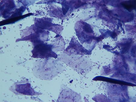
Figure 1. Cytology of an ear swab reveals a mixed population of bacteria and yeast. (All photos courtesy of Dr. Wlson)The breakdown
Diagnostic tests to consider when facing the crusty patient include impression smears, skin scrapes, dermatophyte cultures, aerobic cultures of the skin and multiple punch biopsies for histopathology, Dr. Wilson says. For more superficial bacterial skin infections, like otitis externa, she says that impression smears are the best course of action.
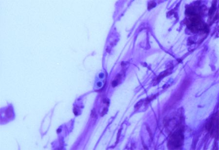
To establish the presence of organisms like bacteria, yeast and fungal spores, smears of pustules and exudative lesions can seriously help (Figures 1 and 2). “Number one, let's establish if this dog has an infection,” she tells attendees.
And if you get a negative result on an impression smear, keep in mind that it could have been a sampling error or any number of common mistakes. “Negative results can be inconclusive,” Dr. Wilson says. “It could still be an infection and you missed it. This can be because we simply missed it on our slide or maybe the owners just gave the pet a bath? Or maybe they forgot to tell you that the white pill they are giving is an antibiotic? Don't be afraid to repeat your cytology if you think you may be missing something. I do it all the time.”
If you're still unsure of the dermatological problem, Dr. Wilson says to take a deep breath and phone a friend. “If you're new or you just don't feel comfortable with it,” she says, “you can always do a slide and send it out to a good clinical pathologist to read it and give you their interpretation. I do cytology constantly, so I'm always looking at impression smears.”

Figure 2. Cytology of an ear swab reveals fungal spores.The process
Dr. Wilson says to first grab your glass microscope slides-“I prefer the ones with the frosted edge,” she says, “because if it doesn't have that, I have no clue which side I did my smear on.” Then you just need your wooden cotton applicators-with or without a heat source for sample fixation-Diff-Quik staining materials, and a microscope with a scanning lens (4x) and an oil immersion lens (100x).
Whether you perform direct impression smears or tape-prep, one slide or multiple slides, Dr. Wilson says it's completely up to personal preference. For her, though, she likes to use one slide per crusty patient. “Technique-wise, you just pretty much take the slide-I'll use a corner of the microscope slide to poke a pustule, I'll use the edge to kind of lift-and then I'll smear all of that on the slide,” she tells attendees.
Sometimes it's a little more difficult to get a slide into a patient's ear, Dr. Wilson says. For that, she recommends a cotton swab on those hard-to-reach areas that are then smeared onto the slide. “I am constantly doing cytology slides,” she admits to attendees, and, eventually, she says, you'll find your rhythm in what process works best for you.
Newsletter
From exam room tips to practice management insights, get trusted veterinary news delivered straight to your inbox—subscribe to dvm360.