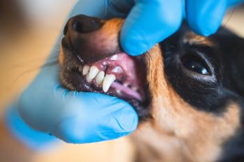
How to repair sick teeth
Extraction isn't always the best choice for veterinary patients with tooth troubles. Find out in which cases tooth repair-not removal-can restore dental health.
When patients present with an illness, we diagnose and treat the treatable. But when they present with sick teeth, the most common dental procedure performed after scaling and polishing is tooth extraction. What can be done to treat and repair dental pathology in our patients when the tooth can be fixed with a reasonable prognosis and the client is willing and able?
In this second article of a series on dental care, we explore repairing teeth with pathology from the outside, inside and surrounding support tissue. While some of the procedures can be performed by general practitioners, others should be attempted only with a full understanding of the anatomy, physiology, procedure and aftercare involved and by individuals with proper equipment and training. Referral to a dental specialist should be considered for advanced dental techniques.
Enamel repair
Enamel, the hardest tissue in the body, protects the underlying coronal dentin and pulp. Enamel disease presentations include enamel hypoplasia, hypomineralization, fracture, abrasion and attrition.
Enamel hypomineralization results from inadequate mineralization of the enamel matrix. The crowns of affected teeth are covered by soft enamel that may be worn rapidly, leaving exposed sensitive dentin (Figure 1A).
Figure 1A. Teeth affected by enamel hypomineralization. (All photos courtesy of Dr. Jan Bellows)
Enamel hypoplasia occurs secondary to disrupted deposition of the enamel matrix between 9 and 12 weeks of age caused by infection, poor nutrition or trauma. Enamel hypoplasia can affect one or several teeth and may be focal or multifocal. The crowns of affected teeth have areas of normal enamel next to areas of hypoplastic or missing enamel (Figure 1B).
Figure 1B. A tooth with enamel hypoplasia. Note the areas of normal enamel next to missing enamel.
Repair consists of applying light-cured acrylic resin over dental bonding agent and adhesive on the prepared areas void of enamel to protect the underlying dentin and pulp. Canines and carnassial teeth are often further restored with metallic crowns to help protect the underlying structures from sensitivity from pressure, heat and cold (Figures 1C and 1D).
Figure 1C. Acrylic restoration of enamel defects.
Figure 1D. Metallic restoration of the crown for maximum protection.
Enamel fractures, attrition (tooth rubbing on another tooth) and abrasion (tooth rubbing on a foreign object) also result in enamel loss. When the dentin is exposed and the dog is young, a similar treatment as that used for enamel hypoplasia is indicated (Figures 2A and 2B). When a mature dog presents with minimal enamel loss, usually no treatment is needed unless dentin or pulp is also exposed.
Figure 2A. Focal loss of enamel due to attrition, exposing dentin in a 2-year-old dog.
Figure 2B. The tooth restored with light-cured acrylic resin.
Repairing inside the tooth
Tooth trauma. A traumatized tooth can present discolored secondary to irreversible pulpitis, with part of the enamel and dentin exposed (uncomplicated fracture), or as a complicated fracture with pulp exposure. Consideration of the tooth's importance, animal's age, age of the fracture and degree of pathology will help you decide if repair is possible.
A discolored tooth usually results from acute trauma causing pulp swelling and internal bleeding against an unyielding pulp chamber. In time, the pulp dies and the tooth becomes nonvital. People with similar conditions often complain of dull pain. If the discoloration is noted acutely with only the coronal tip affected, anti-inflammatory and pain control medication can be used to treat the pulpitis and, hopefully, prevent pulpal death (Figure 3).
Figure 3. A discolored coronal tip, which was treated with anti-inflammatory and pain control medication.
For all other cases of discolored teeth, root canal therapy or extraction is indicated (Figures 4A and 4B).
Figure 4A. A discolored maxillary canine secondary to irreversible pulpitis.
Figure 4B. An intraoral radiograph showing root canal therapy for repair.
Uncomplicated enamel and dentin (near pulp exposure) fractures enter enamel and dentin approaching the pulp. Treatment depends on the animal's age and the fracture's proximity to the underlying pulp. Restoration with acrylic resin covering the exposed dentin is indicated if the animal is young and the pulp cannot be clinically visualized (Figures 5A-5D).
Figure 5A. An uncomplicated enamel and dentin fracture of the right mandibular canine.
Figure 5B. Application of light to polymerize (harden) the acrylic resin.
Figure 5C. Crown preparation before crown delivery.
Figure 5D. Metallic crown restoration.
When the pulp is visualized but not exposed, direct pulp capping is indicated to decrease tooth sensitivity and bacterial invasion through exposed dentin tubules (Figures 6A-6D).
Figure 6A. An uncomplicated crown and root fracture of the maxillary first molar. The underlying pulp is visualized but not directly exposed.
Figure 6B. Application of calcium hydroxide to the pulp.
Figure 6C. Crown preparation for impression.
Figure 6D. The restored tooth with metallic crown.
If the near pulp fracture occurs in an older animal (more than 12 months), there is an increased amount of dentin between the fracture and pulp resulting in less sensitivity. In these cases, you have one of three options:
1. Root canal therapy
2. Serial radiographs (every four to six months) to detect signs of endodontic involvement before root canal
3. Extraction if the client will not agree to root canal therapy or periodic follow-up.
The pulp is exposed in complicated crown fractures. When the fracture is acute (up to two days), vital pulp therapy can be attempted with a guarded prognosis. Extraction or conventional root canal therapy can be performed with a more predictable outcome of an excellent prognosis (Figures 7A-7C).
Figure 7A. An acute complicated canine crown fracture in a cat.
Figure 7B. Application of mineral trioxide aggregate (MTA) during vital pulp therapy.
Figure 7C. Crown restoration with light-cured flowable composite.
When the pulp exposure is chronic, extraction or root canal therapy is the treatment of choice. Leaving a pulp-exposed tooth without treatment allows oral bacteria to enter the patient's blood and create painful inflammation and infection at the tooth's apex (Figures 8A and 8B).
Figure 8A. A complicated crown fracture with pulpal exposure.
Figure 8B. An intraoral radiograph showing root canal therapy to save the tooth with excellent prognosis.
Tooth resorption. In cases of type 2 tooth resorption where the tooth's root is being replaced by bone as confirmed by intraoral radiography, treatment other than an extraction is possible. Crown reduction with gingival closure can be performed to eliminate tooth exposure to the oral cavity and allow continued root replacement resorption. Intraoral radiography is a must to properly identify those cases that would benefit from this procedure (Figures 9A-9D).
Figure 9A. An intraoral radiograph confirming Type 2 tooth resorption.
Figure 9B. Clinical examination of the tooth reveals tooth resorption affecting the mandibular third premolar.
Figure 9C. Crown reduction to treat type 2 tooth resorption.
Figure 9D. Gingival closure after crown reduction.
Dr. Jan Bellows owns All Pets Dental in Weston, Florida. He is a diplomate of the American Veterinary Dental College and the American Board of Veterinary Practitioners. He can be reached at (954) 349-5800; email:
Newsletter
From exam room tips to practice management insights, get trusted veterinary news delivered straight to your inbox—subscribe to dvm360.


