Intraoral films: 7 compelling reasons for every dental patient
According to industry estimates, less than 10 percent of small animal practices have dental radiograph units and of those, less than 10 percent take intraoral films on every dental case.
According to industry estimates, less than 10 percent of small animal practices have dental radiograph units and of those, less than 10 percent take intraoral films on every dental case.
That equates to only 1 percent of the dogs and cats placed under general anesthesia for oral assessment, treatment and prevention (oral ATP) visits get the benefit of survey films.
Reasons given for not taking films include client resistance due to radiation exposure and additional fees, difficulty in exposing and processing films, film interpretation and the added anesthetic time. They are all valid reasons but not insurmountable with proper equipment, dedicated dental radiograph unit and digital sensor, training and client education. In an article published in the Journal of Veterinary Research, Dr. FJ Verstraete et al found that significant dental lesions were noted in 70 percent of the cases in dogs and approximately half of the cats studied.
Hopefully after reading these seven additional reasons to take intraoral films, some of the practitioners not taking dental films routinely will change their minds. All of these reasons came from my general practice from pets presented for teeth cleaning.
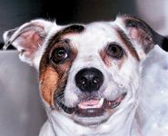
Photo 1a: Dylan
Reason 1: Dylan, a 7-year-old mixed dog, presented for an oral ATP visit. Physical examination showed a moderate amount of plaque and calculus. The right mandibular fourth premolar slightly overlapped the first molar. Full-mouth survey films revealed significant bone loss between the mesial root of the right mandibular first molar and the distal root of the third premolar.
Treatment consisted of exposing the area via gingival flap, curettage and placement of an osteoconductive material (Consil® Nutramax) with a guarded prognosis (Photos 1a, 1b and 1c).
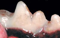
Photo 1b: Dylan's right mandibular fourth premolar and first molar.

Photo 1c: Radiograph revealing significant bone loss between the mandibular fourth premolar and first molar.

Photo 2a: Fred

Photo 2b: Fractured maxillary right second incisor.
Reason 2: During the initial assessment of Fred, a 4-year old Yorkshire Terrier, a right maxillary second incisor fracture was noted. Clinically, pulp exposure was not present. Full-mouth survey films revealed a 3-mm by 3-mm periapical lucency around the right maxillary second incisor extending to the third incisor. The fractured tooth was extracted (Photos 2a, 2b, 2c, 2d and 2e).
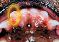
Photo 2c: Fractured maxillary right second incisor apparent near pulp exposure.

Photo 2d: Intraoral radiograph reveals periapical lucency.
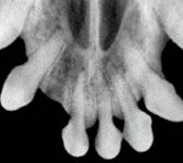
Photo 2e: Post extraction intraoral radiograph.
Reason 3: Marshmello, a 3-year-old Maltese, presented for an oral ATP visit. Assessment under anesthesia revealed slightly mobile right maxillary second and third premolars. Intraoral radiographs revealed deciduous teeth with greater than 75-percent bone loss. The affected teeth were extracted (Photos 3a, 3b and 3c).

Photo 3a: Marshmello
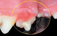
Photo 3b: Right maxillary persistent primary second and third premolars.
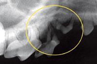
Photo 3c: Intraoral radiograph revealing marked bone loss between the overlapping teeth
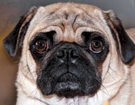
Photo 4a: Puggles
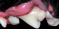
Photo 4b: Gingival recession left maxillary fourth premolar.
Reason 4: Puggles a 5-year-old Pug presented for halitosis. Physical exam revealed gingival recession approaching the mucogingival line of the left maxillary fourth premolar and rotated second and third premolars. Intraoral radiographs revealed almost complete bone loss rostral to the mesial root of the left maxillary fourth premolar as well as support loss of the second and third premolars. The affected teeth were extracted (Photos 4a 4b and 4c).
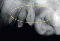
Photo 4c: Radiograph revealing >90 percent bone loss left mesial root of maxillary fourth premolar.
Reason 5: A 2-year-old Basenji, Kody's physical exam under anesthesia revealed slight inflammation and edema surrounding the right mandibular second molar. Intraoral films confirmed bone loss around the furcation as well as >50-percent bone loss around the mesial root. The mandibular second molar was extracted (Photos 5a, 5b and 5c).

Photo 5a: Kody
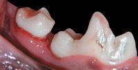
Photo 5b: Right mandibular caudal cheek teeth.

Photo 5c: Radiograph revealing >50 percent bone loss mesial root of the mandibular second molar.

Photo 6b: Right mandibular cheek teeth missing first premolar.
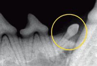
Photo 6c: Radiograph revealing rostrally deviated impacted first premolar.
Reason 6: Max, an 8-month-old Brussels Griffon, presented for neutering. Physical examination under anesthesia revealed a clinically missing right mandibular first premolar. Intraoral radiographs revealed a rostrally impacted first premolar. To avoid a future dentigerous cyst and periodontal disease, the tooth was extracted via flap exposure (Photos 6a, 6b and 6c).

Photo 6a: Max
Reason 7: Buddy, an 8-year-old cat, presented for an oral ATP visit. Physical examination showed minimal inflammation surrounding the left mandibular third premolar. Intraoral radiographs revealed marked internal and external resorption. The tooth was extracted via flap exposure (Photos 7a, 7b and 7c).

Photo 7a: Buddy

Photo 7b: Left mandibular cheek teeth.

Photo 7c: Radiograph revealing marked internal and external resorption.
Dr. Bellows owns Hometown Animal Hospital and Dental Clinic in Weston, Florida. He is a diplomate of the American Veterinary Dental College and the American Board of Veterinary Practitioners. He can be reached at (954) 349-5800; e-mail: dentalvet-@aol.com.

Dr. Bellows