Just Ask the Expert: Dentigerous cysts
Q: How should I treat dentigerous cysts?
Dr. Carmichael welcomes dental questions from veterinarians and technicians.
To ask your question, e-mail: vm@advanstar.com
With the subject line: Dental Questions
Q: How should I treat dentigerous cysts?
A: Although dentigerous cysts are technically nonmalignant lesions, their diagnosis and treatment should not be taken lightly. Dentigerous cysts, which can be very invasive and expansile-even into bone, require thorough surgical removal and curettage.1
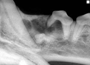
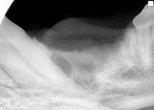
Dentigerous cysts are associated with unerupted or impacted tooth structures; the cysts form when abnormal dental epithelial tissue expands (Figure A). Treatment involves removal of the entire unerupted or impacted tooth structure and thorough curettage of the epithelial lining of the cyst wall. This epithelial debridement, which can be performed with an appropriately sized bone curette, should be as thorough as possible to avoid leaving any remnants of epithelial cyst lining behind (Figure B ). After tooth removal and cyst wall debridement, close the cystic defect; bone remodeling will occur over the course of several months to eliminate the cyst cavity. Provide analgesia for 5 days after surgery.
The key to optimal care is early recognition and intervention before destructive expansion of the cyst occurs. At 7 months, puppies' permanent teeth should have erupted. If not, radiograph each missing tooth to screen for an unerupted or impacted tooth. Extract any unerupted or impacted teeth. Pay special attention to the mandibular first premolar area of small breed, brachycephalic dogs because teeth in this area frequently do not erupt or are impacted. Dentigerous cysts are rare in cats.

Figure A. A dental radiograph of the mandible of a 3-year-old castrated male bearded collie. No evidence of pathology was found on oral examination, other than a missing mandibular first premolar. This radiograph reveals an abnormal unerupted mandibular first premolar associated with radiolucent cystic lesions. Resorption of the mesial root of the mandibular second premolar, caused by the expanding cyst, is also present.

Figure B. A dental radiograph of the patient in Figure A after removal of the abnormal unerupted mandibular first premolar and thorough debridement of the epithelial lining of the cyst cavity. The involvement of the mandibular second premolar necessitated the premolar's extraction as well.
REFERENCE
1. Wiggs RB, Lobprise HB. Clinical oral pathology. In: Veterinary dentistry principles and practice. Philadelphia, Pa: Lippincott-Raven, 1997;131.
Daniel T. Carmichael, DVM, DAVDC
Veterinary Medical Center
75 Sunrise Highway
West Islip, NY 11795
.