Laparoscopy: Underused yet definitive diagnostic tool
The laparoscope was developed as a diagnostic tool in the early 20th Century with the first experimental laparoscopy being performed in a dog in 1901. It wasn't until the 1930s that the laparoscope began being used as a diagnostic tool in human medicine. It took another 50 years before the laparoscope was used to perform surgeries such as appendectomies and cholecystectomies.
The laparoscope was developed as a diagnostic tool in the early 20th Century with the first experimental laparoscopy being performed in a dog in 1901. It wasn't until the 1930s that the laparoscope began being used as a diagnostic tool in human medicine. It took another 50 years before the laparoscope was used to perform surgeries such as appendectomies and cholecystectomies.
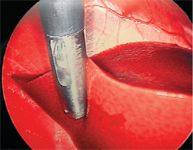
An oval-cup biopsy forceps taking a biopsy from the right lateral liver lobe.
The first documented laparoscopic cholecystectomy in humans was reported in 1985 and within five years became the standard of care. Today, clientele are more educated and aware of the benefits of minimally invasive surgery and are requesting it for their pets. It is incumbent upon the practicing veterinarian to keep up with these technological advances so that they may be offered to their patients.
Veterinary medicine has always lagged behind human medicine, and utilization of the laparoscope is no exception. With all the advanced diagnostic tools available to the practicing veterinarian today, laparoscopy is probably the most underutilized. The advantages of laparoscopy are that it is minimally invasive, yet highly accurate, and can provide definitive diagnostic and staging information. By virtue of its small surgical incisions, there is less physiologic stress to the patient, less pain and a quicker recovery. It is generally accepted that laparoscopic biopsies are superior when compared with tissue samples obtained via other percutaneous methods (i.e. ultrasound-guided needle biopsies). Because the organs are directly visualized and magnified by the laparoscope, smaller lesions (<0.5 cm) that may be missed with other imaging modalities may be detected and biopsied. This type of information can be of vital importance when staging neoplastic diseases and formulating treatment plans. Laparoscopy also carries a very low complication rate (<2 percent). The disadvantages of laparoscopy include the lack of tactile feedback, limited field of vision and inability to completely explore the entire abdomen. In many cases, it does not replace the need for a conventional laparotomy. In addition, laparoscopy requires new surgical skills, formal training and a substantial investment in specialized surgical equipment.

Suggested reading
The most common indications for diagnostic laparoscopy is to visually inspect and biopsy abdominal organs or masses. This allows for accurate staging of neoplastic processes so that appropriate treatment plans can be implemented. The liver, pancreas, spleen, lymph nodes, adrenal glands, kidneys and abdominal masses are all amenable to laparoscopic biopsy.
Intestinal biopsies are possible with laparoscopic assistance but require that the intestinal loop be exteriorized through a small incision and standard incisional biopsies are then taken. The laparoscope can be used also to perform diagnostic procedures such as splenoportography, cholecentesis for bile culture and cholecystography.
The relatively few contraindications to laparoscopy are septic peritonitis, diaphragmatic hernia and cases where conventional laparotomy is indicated. If there is a tear in the diaphragm, the creation of a pneumoperitoneum results in life-threatening tension pneumothorax. Limitations for laparoscopy include small patient size (<2 kg), very obese patients and abdominal effusions.
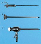
Examples of laparoscopic instrumentation: A) Veress insufflation needle used to establish pneumoperitoneum. B) Trocar. C) Cannula with insufflation port and one-way valve.
Procedural considerations
Excessive falciform fat or abdominal effusions can interfere with accurate visualization. In patients with abdominal effusions, as much fluid as possible should be withdrawn before insufflating the abdomen.
The basic instrumentation required for diagnostic laparoscopy includes a 5 mm, 0-degree field-of-view telescope, 2 cannula/trocar units, Veress insufflation needle, xenon light source and cable, insufflator, CO2 source and various instrumentation. A videocamera/monitor is ideal and is now considered standard when purchasing new equipment.
When purchasing equipment, it is important to consider compatibility with other instruments, ability to expand with other scoping procedures, warranties and technical support.
A complete and thorough work-up is required before considering laparoscopy. Abdominal ultrasound is almost always performed prior to laparoscopy. The ultrasound is helpful in determining location, size and consistency of parenchymal or abdominal lesions.
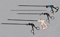
Basic instrumentation used to manipulate and biopsy tissues. Listed from top to bottom: Babcock forceps, palpation probe, scissors, grasping forceps and oval-cup biopsy forceps.
Ultrasound can detect the presence of deeper parenchymal lesions not apparent to the laparoscopist. In cases with liver disease, ultrasound also helps to predict the patency of the biliary system. If a complete biliary extra-hepatic obstruction is suspected, a conventional laparotomy is indicated rather than laparoscopy. Finally, in cases with renal disease, ultrasonography and/or IVP studies may be helpful in detecting changes in renal architecture, the presence of ureteral obstruction, and determining if the disease is uni-or bilateral.
Anesthetic and operative considerations include many of the same guidelines ascribed to traditional surgery. The patient should be fasted for 12 hours to ensure that the stomach is empty. The bladder should be expressed to improve visualization and prevent accidental injury when the cannulas are placed. The operating table should be adjustable so that the patient's position can be changed in order to improve visualization.
Anesthesia needs to be carefully monitored and ventilation assisted because of the pressure that the pneumoperitoneum places on the diaphragm. Finally, the entire ventral abdomen should be prepped in case there is a need to switch to a traditional laparotomy.
The approach
There are three basic approaches to laparoscopy: ventral midline, right lateral and left lateral. The ventral midline approach provides good visualization of the liver, gall bladder, pancreas, stomach, intestines, reproductive system, urinary bladder and spleen. The falciform fat may interfere with visualization with this approach especially if the patient is obese. The right-lateral approach is preferred for liver, gall bladder, pancreatic, duodenal, R-renal or R-adrenal assessment.
About 85 percent of the liver can be visualized from the R-lateral approach. The left-lateral approach is used less frequently because of the proximity of the spleen and the increased chance of iatrogenic splenic damage.
The basic principle of laparoscopy is to create a pneumoperitoneum so that a good viewing window can be established for the telescope. The placement of two or more cannulas or ports allows the telescope and instruments to be manipulated through the abdominal wall. The specific steps of abdominal insufflation and cannula placement can be found in the list of suggested reading.
Laparoscopy is the preferred method of obtaining liver biopsies. A right-lateral or ventral approach allows good visualization of the liver, but the right-lateral approach provides the best view of the biliary system and right limb of the pancreas. When assessing the liver, color, texture, lobular pattern, margins, size and any nodular changes should be noted. Each lobe should be lifted with the palpation probe to examine the dorsal surface of the liver. The biliary system should also be assessed to rule-out obstructive lesions. Bile can be aspirated with a 20g spinal needle if indicated for culture and sensitivity. For biopsies of the liver parenchyma, an oval-cup biopsy forceps is used to obtain samples from at least three to four sites. It is important to biopsy both normal and abnormal-appearing tissue. Even with abnormal coagulation profiles, bleeding is usually minimal. If bleeding is deemed excessive, direct pressure can be applied to the biopsy site with the palpation probe or a piece of saline-soaked gel foam can be applied to the biopsy site. The tissue samples submitted for histopathology should include a thorough history, visual findings and special requests for staining (i.e. for copper or amyloid, if indicated).
Pancreatic biopsies can be of great value in confirming chronic pancreatitis in cats or identifying pancreatic masses such as insulinomas. Despite being taught that the pancreas should not be touched, biopsies of the pancreas can safely be obtained through laparoscopic guidance. A right-lateral approach is used to inspect the right limb of the pancreas.
Visualization of the left limb of the pancreas is difficult. Only one or two samples should be taken from the edge of the right limb of the pancreas, taking care to avoid the pancreatic ducts and vessels. A punch-type biopsy instrument is preferred for the pancreas.
Renal biopsies taken under laparoscopic guidance can yield excellent tissue specimens for histopathology. Renal cysts, hydronephrosis and ureteral obstruction are all contraindications for renal biopsy. Pre-operative diagnostics such as an abdominal ultrasound or IVP should be performed pre-laparoscopy and will dictate which kidney should be biopsied. If there is no preference, the right kidney is chosen because it is less mobile. Care must be taken to avoid penetrating the diaphragm with the biopsy needle because it could result in a pneumothorax. The cranial or caudal pole of the kidney should be targeted to obtain mostly cortex.
The corticomedullary junction should be avoided because of the larger arcuate vessels. A recent study showed that a 14g automatic, core-type biopsy needle yielded better tissue specimens with less crush artifact and more glomeruli than a similar 16g needle biopsy.
With the laparoscope, post-biopsy hemorrhage can be monitored and controlled by applying direct pressure with the palpation probe.
The reported complication rate for laparoscopy is low (<2 percent) but proper training is essential to maintaining that type of success. Operator inexperience can greatly increase the complication rate. There are many good courses taught at major conferences such as the ACVS Symposium and the North American Veterinary Conference. In addition, some universities, such as Colorado State University, offer various scoping courses year round.
Diagnostic laparoscopy is a great way to get started in minimally invasive surgical techniques. Once proficient, the surgeon can then expand the use of the laparoscope to perform various surgical procedures such as laparoscopic-assisted gastropexy, cryptorchid castration and ovariohysterectomy.
Katharine R. Salmeri, DVM, Dipl. ACVS, was a soft-tissue surgeon at Animal Medical Center before joining Red Bank Veterinary Hospital in 1991. She completed a residency in small animal surgery at the University of Florida in 1989 and is a veterinary graduate of Colorado State University.
Podcast CE: A Surgeon’s Perspective on Current Trends for the Management of Osteoarthritis, Part 1
May 17th 2024David L. Dycus, DVM, MS, CCRP, DACVS joins Adam Christman, DVM, MBA, to discuss a proactive approach to the diagnosis of osteoarthritis and the best tools for general practice.
Listen
