Last in a Two-Part Series: Uncovering the pruritic dog takes more than scratching the surface
As mentioned in the first article (Feb. 2002) of this series, the presentation of the pruritic dog can be frustrating for the veterinarian because of the number of possible differential diagnoses.
As mentioned in the first article (Feb. 2002) of this series, the presentation of the pruritic dog can be frustrating for the veterinarian because of the number of possible differential diagnoses.
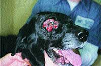
An example of a focal pericular deep pyoderma.
It cannot be emphasized enough that a detailed history and thorough physical examination is essential in determining the cause of the pruritus. A detailed history includes age, breed and sex of the affected animal as well as duration of the pruritus, areas of the body involved, and response to any medications either topical or systemic that have been administered.
A thorough physical examination should include dermatologic procedures such as combings, skin scrapings and cytology of any lesions present. Depending upon the outcome of these in-house tests, other laboratory procedures such as complete blood counts, serum profiles, thyroid panels, tests for Cushing's disease, allergy testing or performing skin biopsies may be necessary. In part one, differentials of pruritus that included ectoparasites were discussed. Now that in-house testing and/or therapy to rule out this category have been performed, the next step is to rule out bacterial pyoderma (primary or secondary), atopy and/or food allergy as the possible cause of pruritus.
Bacterial pyoderma
In the canine, bacterial pyoderma, the majority of the time, is caused by Staphlococcus intermedius. That is why many veterinarians do not feel the need to routinely culture a bacterial pyoderma. However, the time when a culture and sensitivity is necessary is when the patient is not responding to an appropriate antibiotic (assuming antibiotic only is used, not in conjunction with steroids) or when a deep pyoderma is present, particularly, when demodicosis is involved.
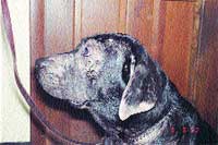
Labrador Retriever with inhalant allergy. The other differential would be scabies because of the ear-edge involvement.
In the latter, many types of bacteria may be present including Pseudomonas and Proteus species. The diagnosis of bacterial pyoderma is usually made by clinical presentation, i.e. the appearance of pustules or epidermal collarettes.
When intact pustules are present, cytology should be performed revealing results consistent with bacterial pyoderma such as degenerative neutrophils with or without intracellular cocci. The main differential for bacterial pyoderma is pemphigus foliaceus which, at times, may be difficult to differentiate for both the clinician and pathologist. In older patients, epitheliotropic lymphoma may appear clinically similar to bacterial pyoderma. A skin biopsy is necessary to differentiate between the two diseases.
Antibiotic therapies
In a "first time" bacterial pyoderma, antibiotics such as sulfa derivatives (use with caution in patients with keratoconjunctivitis sicca (KCS), those breeds prone to KCS, or in Dobermans which can show a possible genetic predisposition for adverse reactions to sulfas), cephalosporins, macrolides such as lincomycin, or b-lactamase potentiated amoxicillin (Clavamox) should be administered until total clearing of the pyoderma plus an additional week past clearing. Antibacterial bathing should also be performed.
Tetracyclines and penicillins normally are not effective in treating canine staph pyoderma. Steroids should be avoided even if the patient is pruritic as they may cause immunosuppression thereby "undoing" the effects of the antibiotic. Normally 21 to 30 days of antibiotic is initially dispensed with a recheck at the end of that period of time to be sure the pyoderma is resolving, then additional antibiotic is dispensed to complete the "one week past clearing" process. Antibacterial shampoos such as chlorhexidine, ethyl lactate (Etiderm), benzoyl peroxide or sulfa/salicylic acid combinations can be helpful adjunctive therapy. In a recurrent pyoderma, factors to consider include: Was the pyoderma treated with an appropriate antibiotic without steroids until one week past clearing? Is there a potentiating underlying disease such as demodicosis, hypothyroidism, Cushing's disease, diabetes mellitus, atopy or food allergy? Treatment of a recurrent pyoderma involves antibiotics, antibacterial bathing and addressing the underlying disease perpetuating the pyoderma.
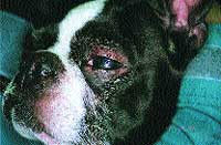
Periocular alopecia and erythema in an atopic Boston Terrier.
If no underlying disease is found, then "pulse dosing" antibiotics i.e. one week on/one week off is performed or immune stimulants such as Staphage Lysate are administered.
Dietary troubles
Food allergy can manifest with many symptoms including a recurrent bacterial pyoderma, otitis, Malassezia dermatitis and pruritus without lesions. Dogs of any age, breed or sex can be affected. Some dermatologists classify food allergic dogs as having symptoms involving "ears and rears".
Nondermatologic manifestations of food allergy can include gastrointestinal problems such as vomiting, diarrhea, and flatulence, respiratory problems or neurologic symptoms including seizures. Food allergic patients usually have nonseasonal problems since they are eating the same food all of the time (or variations of the same food) and are nonresponsive to antipruritic doses of steroid (however some patients may get temporary relief from anti-inflammatory or immunosuppressive doses of steroid).
Since blood or skin testing for food allergy has not been proven to be accurate, the best way to assess food allergy in the dog is the feeding of a hypoallergenic diet either cooked or commercially prepared for at least eight to 10 weeks and perhaps longer. The favored method is a home-cooked diet consisting of a novel protein and single carbohydrate source.
Since this is impractical for most large breed dog owners, commercially available prescription foods containing novel proteins such as venison, rabbit, duck, fish, kangaroo or a protein hydrolysate diet are available. The concept of what one is trying to accomplish must be explained to the owner so that the diet is performed correctly.
The point is to remove the pet from all ingredients to which they have been exposed to in previous foods or treats e.g. corn, wheat, egg, beef, chicken, soy and dairy. No other foods, bones, rawhides, treats or flavored heartworm preventatives should be administered during the diet period.
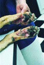
Erythemic alopecia caudal in an atopic patient.
Some owners elect to administer distilled water as opposed to tap water to further control what the animal is receiving. It may be difficult to undertake a hypoallergenic diet for owners with young children since theoretically a dietary indiscretion can set the patient back one to two weeks. Since there are two categories of hypoallergenic diets, the single allergen diet and the protein hydrolysate diets, a patient may respond to one type of diet and not the other. If you have undertaken a hypoallergenic diet of one particular type without success, be sure to feed one of the other category to fully rule out food allergy. Be extremely inquisitive about what is administered to the patient i.e. medications cannot be given with cheese, lunch meats or peanut butter (contains corn syrup) which will negate the effect of the diet.
Inhallant allergy
Atopy or inhalant allergy affects 3-15 percent of dogs. It seems that atopic patients are showing signs of atopy including pruritus without lesions and recurrent pyoderma at younger and younger ages. It is not unusual to see some patients at 4 to 6 months of age manifesting early signs of atopy. Age of onset is usually between 6 months and 3 years of age but occasionally the middle-aged to older dog may appear with symptoms. Common breeds affected include Retrievers, Dalmatians, Dobermans, Shepherds, herding breeds, Chows, Shar Peis and terriers.
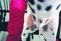
Groin erythema from chronic licking in a food allergic Dalmatian.
Symptoms may be seasonal or nonseasonal depending upon what they are allergic to. Patients allergic to house dust mites may have problems year round, whereas those with seasonal pollen allergies are affected only during a particular season.
"Inhalant" allergy denotes the inhalation of the allergen resulting in the body's reacting to that allergen, however some evidence in humans suggests a percutaneous absorption of the allergen. In the dog this would involve the nonhaired areas of the body such as the feet, groin and ventral abdomen. Most inhalant allergic dogs tend to be flea allergic and harbor Malassezia and/or bacteria. Keeping the secondary yeast and/or bacteria under control can be a major part of treating atopy in the dog successfully.
Most atopic patients tend to respond to antipruritic doses of steroid (e.g. prednisone 0.25mg/lb/day). However steroid tolerance can be a problem with prolonged use of steroids as well as their undesirable side effects. For those reasons we all strive to control atopy with a minimal use of steroids.
Frequent bathing in an antipruritic shampoo, antibacterial shampoo or just a mild cleansing shampoo is helpful in keeping the bacterial counts reduced as well as mechanically removing the allergen, particularly in pollen allergic patients.
Fatty acids either via the diet or oral capsules/granules may be helpful in producing less inflammatory mediators of inflammation. Fatty acids in combination with antihistamines may produce a higher success rate used together than when either is used alone. Remember to try each of the different categories of antihistamines before deeming this therapy as unsuccessful.
Once the diagnosis of atopy has been made by breed, history, age of onset and response to therapy, skin or blood testing to see what allergens need to be included in the patients allergy solution may be undertaken. Do not use invitro testing to make the diagnosis of atopy, just to know what to include in the patient's allergy solution. Whether the patient is blood or skin tested, the results must concur with the season the patient is affected.
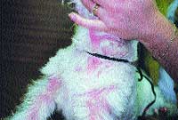
Generalized erythema in a food allergic patients with secondary malassezia yeast dermatitis.
Immunotherapy is approximately 65-70 percent effective in controlling symptoms of atopy but may take up to a year to become effective. Other medications such as cyclosporine and pentoxifylline have been used in dogs for atopy. Side effects of oral cyclosporine at 5mg/kg/day include vomiting and since renal dysfunction can be a problem in humans, monitoring serum creatinine and urinalyses in the dog is suggested. Long-term effects of cyclosporine when used for atopy in the dog are unknown.
Treatment
It must be remembered that some patients may be atopic and food allergic. If you feel you have relieved a patient's symptoms with some degree of success but you just can't get them "perfect", the patient may have two problems. In atopics, it may be prudent to undertake a food trial in addition to immunotherapy for inhalant allergy. The breed we see most commonly affected with food and inhalant allergy is the Labrador Retriever. For example, the recurrent pyoderma and pruritus may be relieved by getting the atopy under control, but the otitis remains a problem. A hypoallergenic food trial may resolve the otitis you can't seem to get under control.
Finally, if a patient continues to respond to antibiotics with resolution of the lesions and the pruritus and food and inhalant allergy have been ruled out, bacterial hypersensitivity is a differential. Therapy includes "pulse" dosing of antibiotics i.e. one week on/one week off the full dose of antibiotics or immune stimulants such as staphage lysate. Staphage lysate injections are administered twice weekly up to a dose of 0.5cc twice/week.