Malocclusion with severe tissue destruction in a dog
Learn about the multiple options available to treat this condition.
Last month, I began a discussion on the topic of malocclusions. Over the next several months, I will take a close look at cases of malocclusion resulting in varying degrees of trauma and how they were treated. There is generally more than one option for treating each of these conditions, and alternative techniques will be addressed.
Signalment and oral examination findings
A 28-month-old intact male Doberman pinscher presented with linguoversion of the right mandibular canine tooth (404). The left mandibular canine tooth (304) occluded normally. Tooth 404 had created an oronasal fistula and destroyed the palatal and vestibular mucosa adjacent to the right maxillary canine tooth (104) (Photo 1).
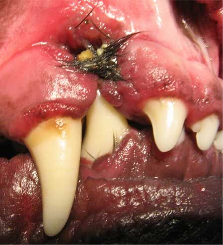
Photo 1: Tooth 404 destroyed the palatal and vestibular mucosa adjacent to the right maxillary canine tooth and created an oronasal fistula.Hair, food debris and pus were present within the defect (Photo 2). The American Veterinary Dental College describes this condition as a class I malocclusion or neutroclusion. According to this description, there is a normal rostrocaudal relationship between the mandibular and maxillary arcade but with malposition of one or more teeth.
Although palatal trauma is common with this condition, most patients that present with linguoversion of one or both mandibular canines do not demonstrate such profound tissue destruction.
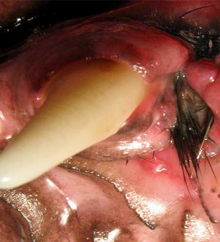
Photo 2: Hair, food debris and pus were seen within the defect.Therapeutic options
One possible option for resolving this malocclusion is creating a bilateral incline plane to passively move tooth 404 into the diastema between the right maxillary third incisor (103) and tooth 104. This option poses some limitations and possible complications. A Mann incline plane is a fixed cast appliance that is anchored to the maxillary canine teeth.1 This option requires a minimum of three anesthetic episodes-one to create alginate impressions and stone model creation for laboratory fabrication of the device, one to place the device and a third to remove the device. The chance of this appliance being dislodged is extremely low. It is the best alternative in a large adult patient with this malocclusion if an incline plane is chosen for therapy.
An alternative incline option can be fabricated directly using acrylic (Photo 3). This option requires anesthesia both for the placement and removal of the appliance. The breakdown of the acrylic, especially in large patients, is a potential complication. Although this appliance requires one less anesthetic episode, because of the potential impact of premature breakdown on the soft tissue defect, it is not a good option for this patient.
Extracting tooth 404 would eliminate the malocclusion and is a viable alternative. Advanced surgical extraction skills are required to eliminate potential complications of fracture or mucoperiosteal flap dehiscence if extraction is elected. Time, expense and potential complications are drawbacks of this technique.
One consideration for resolution of this malocclusion-and the option chosen in this case-is crown reduction, partial pulpectomy and vital pulp therapy.2 Minimal anesthetic time, decreased owner expense and immediate resolution of the malocclusion are benefits to this approach. Referral to a veterinary dental specialist is recommended for this procedure. It is technique-sensitive and requires proper training and specific materials for optimal success. Although this procedure is highly successful when performed correctly, monitoring the tooth radiographically is recommended to document continued viability.
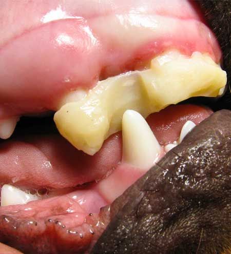
Photo 3: An acrylic incline plane, as seen here, was not the best option for the patient in this case. Therapy
Although this article is not designed as a step-by-step reference for crown amputation, partial pulpectomy and vital pulp therapy, the following is a brief discussion of the technique.
After cleaning and polishing the tooth, a disinfection solution is applied to minimize microbial contamination. A sterile cross-cut tapered fissure bur is used to amputate the crown at the level of the incisor teeth, and 5 mm of vital pulp tissue is then carefully removed by using a dental bur. I prefer a fine-tapered diamond bur since it minimizes tearing of the pulp and decreases hemorrhage. Moist paper points are gently applied to eliminate hemorrhage, and pulp dressing consisting of mineral trioxide aggregate or calcium hydroxide is placed directly on the pulp (Photo 4).
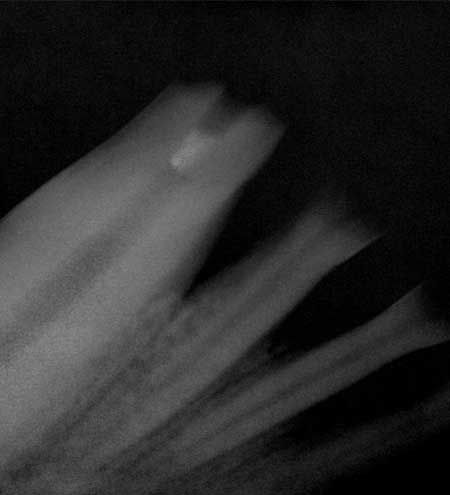
Photo 4: By using a bur, 5 mm of pulp was removed. Mineral trioxide aggregate can be seen as the radiodense material within the pulp cavity.A glass ionomer and final composite layer followed by two applications of an unfilled resin complete the procedure (Photos 5 and 6).
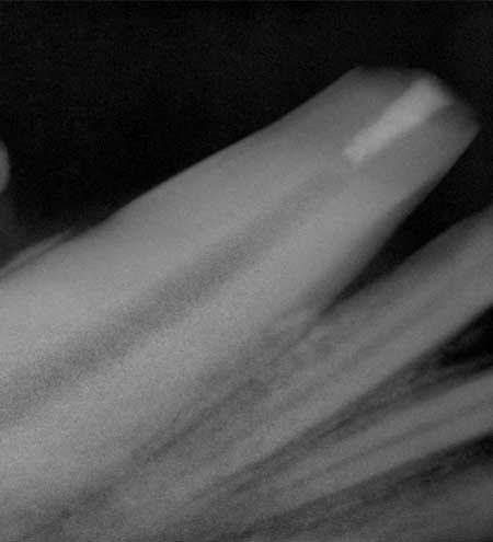
Photo 5: A glass ionomer and final composite layer followed by two applications of an unfilled resin completed the procedure.
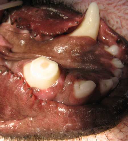
Photo 6: This photo shows the final appearance of tooth 404 after crown amputation, vital pulpectomy and vital pulp therapy.Unfortunately, in this patient, eliminating the source of the defect did not provide definitive resolution. Débridement and freshening of the edges of the soft tissue defect were performed. Metzenbaum scissors were used to create a mucoperiosteal flap from adjacent tissue sufficient to provide closure without tension (Photo 7).
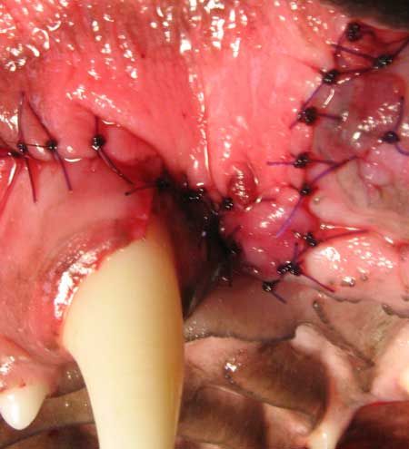
Photo 7: Metzenbaum scissors were used to create a mucoperiosteal flap from adjacent tissue to provide closure without tension.
Despite premature removal of an Elizabethan collar, the patient returned at the three-week recheck demonstrating complete healing (Photo 8).
Conclusion
Multiple options exist for correcting this malocclusion. It is important that clients are aware of all options. Variable presentations occur with similar soft tissue trauma and always necessitate evaluation of the different therapeutic options. One such presentation will be the topic of the next case in this series.
REFERENCES
- Wiggs RB, Lobprise HB. Basics of orthodontics. In: Veterinary dentistry principles and practice. Philadelphia, Pa: Lippincott, 1997;456.
- Mulligan TW, Niemiec BA. Endodontic treatment of vital pulp tissue. Clin Tech Small Anim Pract 2001;16(3):159-167.
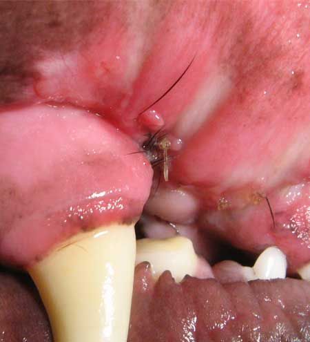
Photo 8: At the three-week recheck, the patient demonstrated complete healing.
Delineating the problem
Read the first article in this multiple-part series, “Defining dental malocclusions in dogs,” and other dental articles by Dr. Beckman at dvm360.com/beckman.

3 ways to help patients smile
The CVC in Washington, D.C., May 4-7, offers these three hands-on wet labs for practicing your dental techniques
- Dental Radiography: Discover the nuances of patient positioning and obtaining, processing and interpreting dental radiographs.
- Dental Techniques for Technicians: Brush up dental cleaning and polishing, charting, hand scaling, and more.
- Practical Dental Techniques: Learn the latest on managing periodontal disease, performing difficult extractions, repairing oronasal fistulas and more.
- To find our more about these labs and to register to attend, visit thecvc.com.