Managing a 9-month-old kitten with FIP
A case study of a neutered male domestic shorthaired cat who presented for evaluation of hyporexia and labored breathing of three weeks' duration.
Sully, a 9-month-old 6.4-lb (2.9-kg) neutered male domestic shorthaired cat, was presented for evaluation of hyporexia and labored breathing of three weeks' duration.
History
Sully was adopted from a shelter at 3 months of age and had negative results for feline leukemia virus (FeLV) and feline immunodeficiency virus (FIV) at the time of the adoption. Sully was current on core vaccines at presentation and was kept exclusively indoors.
Physical examination and routine laboratory findings
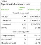
Sully's respiratory rate was 60 breaths/min. Its temperature (102 F [38.9 C]) and pulse rate (200 beats/min) were normal. Heart and lung sounds were muffled on the left side and clearly audible on the right. Significant complete blood count and serum chemistry profile findings are listed in Table 1. FeLV and FIV test results were negative.

Table 1: Significant laboratory results
Diagnostic imaging
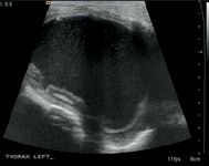
A radiographic examination showed fluid or a soft tissue density in the left hemithorax (Figure 1). A thoracic ultrasonographic examination showed a large fluid-filled cavity in the left hemithorax, bounded by a 3-mm distinct capsule (Figure 2). No fluid was seen in the mediastinum or the right pleural space.

1. A ventrodorsal thoracic radiograph. Note the fluid or soft tissue density on the left side of the thorax. The cardiac silhouette is displaced to the right.
Fluid analysis and PCR testing
A thoracocentesis was performed, and 120 ml of thick straw-colored fluid were removed from left side of the chest. Analysis of the yellow, slightly turbid fluid showed a total protein concentration of 4.5 g/dl, an albumin:globulin ratio of 0.45, and a nucleated cell count of 1,471/μl with primarily nondegenerate neutrophils. The results of PCR FIP mRNA tests performed on whole blood and pleural fluid were negative.

2. An ultrasonographic image of the left hemithorax. Note the thick-walled cystic structure. (This image is courtesy of the Texas A&M University Radiology Service.)
Thoracotomy
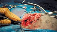
A thoracotomy was performed, and a cystic lesion was found to occupy the left side of the chest. It was adhered to the parietal pleura and a section of atelectatic or malformed lung. Both the cystic lesion and attached lung tissue were removed (Figure 3).

3. An intraoperative view of the left hemithorax. The cystic structure and affixed lung tissue are visible, following their removal from the chest.
Histology
The tissue labeled cyst was a capsule composed of smooth muscle and granulation tissue surrounding fibrinonecrotic and suppurative inflammation. The tissue labeled lung was atelectatic lung with a capsule of smooth muscle and granulation tissue surrounding fibrinonecrotic and suppurative inflammation. Special stains for bacteria and fungi were negative.
Immunohistochemistry
The portions of the tissue submitted for immunochemistry were positive for the FIP antigen, which confirmed the diagnosis of FIP.
Outcome
Sully was treated with glucocorticoids (prednisolone 2 mg/kg orally once a day). It did well until three months after the surgery, when the cat developed pericardial effusion. Sully was euthanized about four months after the thoracotomy. At necropsy, fibrinous plaques were noted on the pleura and pericardial sac and covering the abdominal viscera.
Comments
This case is unusual because FIP is not commonly associated with a unilateral pleural effusion. Although there was strong suggestive evidence of FIP on the basis of the fluid analysis, mature neutrophilia, and hyperglobulinemia, the owner was determined to establish a definitive diagnosis and opted to pursue a thoracotomy. It is interesting to note that mRNA PCR test results were negative, despite the final diagnosis of FIP. This case highlights the challenges associated with this disorder and the limitations of noninvasive diagnostic tests.