Nasal parasites and their differentials In the dog and cat (Proceedings)
Parasites are minor cause of nasal disease in dogs and cats. However, they should be added to a differential diagnosis list of nasal disease. This review will discuss the biology, diagnosis, disease, and treatment of these parasites, and discuss the differential diagnosis, and the methodology for treating at least one differential diagnosis, that of nasal aspergillosis in dogs and cats.
Parasites are minor cause of nasal disease in dogs and cats. However, they should be added to a differential diagnosis list of nasal disease. This review will discuss the biology, diagnosis, disease, and treatment of these parasites, and discuss the differential diagnosis, and the methodology for treating at least one differential diagnosis, that of nasal aspergillosis in dogs and cats.
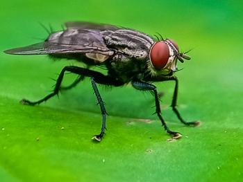
1. Cuterebra spp.
Epidemiology: Arthropod dipteran parasite; some 34 different species in North America.
• Large maggots seen in the skin of dogs and cats and represent the larva of the rodent and rabbit bots. Adult flies lay eggs around the entrances to rodent burrows, and when host passes, the small first-stage maggot hatches and jumps onto the passing host.
• Larva capable of entering the host through the mouth, nose, eyes, or anus.
• In rodent hosts, larvae remain as 1st-stage larvae within the nasopharyngeal region near the posterior end of the soft palate and in the nasal passages. Maturation time from infection to when the larva leaves the warble is between 3 to 8 weeks.
• Seasonality: Adult flies have single emergence in late spring in the more temperate parts of the United States.
• Cats: Infected as they hunt. Kittens may be infected by larvae brought back on the fur of the queen.
• How larvae enter cats unknown, but probably through the mouth, nose, or anus as they do in the rodent and lagomorph hosts.
Clinical signs: Depend mainly on where the larva locates.
• Skin lesions; (warble) containing a single larvae usual present on the cheek, neck, or back, but other sites are reported.
• Acute upper respiratory tract distress; severe sneezing, unilateral initially serous then mucopurulent nasal discharge. Sneezing and nasal discharge can persist for a week.
• May be accompanied by unilateral facial swelling especially over the nose.
• Bloody nasal discharge, soft palate and pharyngeal swelling have been reported.
• Laryngeal edema (larval migration in the cervical neck) may cause laryngeal edema and arrest. Respiratory signs may persist for few days to several weeks, then often recede in severity occasionally to be followed by neurologic signs
• If neurologic signs develop, they will do so one to two weeks after respiratory signs, although respiratory signs have been reported to occur as long as 4 to 10 weeks before the onset of neurologic signs.
Diagnosis: Viewing the larvae within the respiratory tract, or made on circumstantial evidence of acute rhinitis (sometimes progressing to neurologic disease) in an outdoor cat during late summer and fall. The larvae or its migration tracts through the brain may be identified on CT scan or MRI.
Treatment: Ivermectin (0.1 to 0.3 mg/kg, PO, q24h, 3 days) very effective.
• Prednisone (1 mg/kg, PO, q12h, 3 weeks, then 1 mg/kg, PO, q24h, 3 weeks). [Diagnosis based on clinical signs, but usually improve). Few develop neurologic disease. Some neurologic cats improve clinically with this treatment but outcome unchanged.
2. Pneumonyssoides caninum (canine nasal mite)
Epidemiology: Arthropod parasite of nasal sinuses of dogs. Occurs in the USA, Canada, Japan, Australia, Sth. Africa and Europe.
• Adult mites (1 mm X 0.5 mm in size) are easily identified by their leg morphology; the first pair of legs each terminate in a pair of large hooks, while legs two, three, and four each terminate in a sucker armed with a pair of smaller hooks.
• Believed that dog-to-dog transmission is by the direct transfer of larvae from one infested dog to another.
Clinical signs: Sneezing, although can present with facial pruritis, snuffling, snorting ± nasal discharge and excessive lacrimation.
Treatment: Ivermectin (200 μm/kg body weight, PO or SC) or Milbemycin (0.5 mg/kg, PO) once weekly for 3 weeks are effective.
3. Linguatula serrata.
Epidemiology: Pentastomid parasites that represent a group of specialized crustacean-like arthropods.
• Adult female is approximately 8 to 10 cm long and 1 cm in diameter; the male is about 2 cm long. The body appears superficially annulated, and the worms tend to be tan to brown in color.
• Life cycle requires an intermediate host. The eggs that are passed by the female contain a four-legged larvae. Eggs do not appear in the feces of the dog but instead are found in the nasal secretions
• Suspected that most dogs obtain their infections by the ingestion of sheep offal. When dogs ingest infected tissues, the nymphs migrate up the back of the throat into the nasal turbinates. Once swallowed, the nymphs do not migrate back up the esophagus.
• Prepatent period (PPP) is about 6 months. Adult worms live about 2 years.
Clinical signs: Sneezing, slight nasal discharge sometimes containing blood. The parasites become large, lie in the recesses of the nasal turbinates, and attach themselves firmly to the mucous membranes with their four hook. The adults apparently feed on respiratory mucosal cells and blood. When fully grown, the parasites are capable of causing nasal obstruction.
• Humans may also become infected but not from dogs.
Diagnosis: Eggs (yellowish oval, 80 Φm, surrounded by bladder-like envelope and containing a four-legged larva) in the nasal secretions. - identifying larvae during rhinoscopy.
Treatment: Physical removal only treatment described. Ivermectin (200 μg/kg, PO, once) may be efficacious.
4. Eucoleus boehmi.
Epidemiology: Capillarid nematode parasite of nasal mucosa of the dog. Adult worms live threaded through the mucosa of the nasal sinuses. Adults appear as very fine threads seen grossly as very fine transparent hairs.
Clinical signs: Sneezing, nasal discharge.
Diagnosis: Identifying eggs in feces (eggs can be recovered in nasal washings).
Treatment: Fenbendazole (50 mg/kg, PO, q24h, 7 days). Ivermectin (200 μg/kg, PO, once).
DIFFERENTIAL DIAGNOSIS
1. Neoplasia
2. Tooth root abscess
3. Viral rhinitis (cats)
4. Fungal rhinitis
5. Foreign body
6. Allergic rhinitis
Although each of these conditions presents therapeutic challenges, improvement in the methodology of treating fungal rhinitis has resulted in marked improvements in the outcome of this condition.
5. Fungal Rhinits (Nasal Aspergillosis)
Diagnosis
• Serum biochemistry and CBC are non-specific - neutrophilic leukocytosis, monocytosis.
• Nasal radiographs – increased radiolucency in affected rostral and maxillary nasal turbinate area (turbinate lysis) especially on open-mouth ventrodorsal and skyline views of frontal sinuses; mixed densities seen due to turbinate destruction and soft tissue dense fungal granulomas or accumulated nasal discharge.
• CT and MRI – accurately define extent of disease and allow assessment of the integrity of the cribiform plate; important when considering treatment with local infusion of anti-fungal agents.
• Rhinoscopy – directly visualize fungal granulomas; appreciate destruction of nasal turbinates (cavernous appearance in severe cases); collect material for culture, cytology, and histopathology.
• Fungal culture – should be taken from specific lesions or may give false positive results as the organism is a common contaminant.
• Positive cultures should be confirmed by histopathology or cytology.
• Serology (agar gel double diffusion – AGDD and counterimmunoelectrophoresis CIE) – ELISA lacks accuracy and should not be used: – supports a diagnosis if made in conjunction with culture and evidence of disease (consistent radiographic changes in nares); the AGDD is high sensitive and specific; may get false negatives early in infection; titers often remain elevated in dogs successfully treated for nasal aspergillosis so serology is not a good way of monitoring the outcome of therapy.
• False positives and cross-reactivity with Penicillium spp. reported; however, recent tests may miss Penicillium spp. as antigens used in tests often very specific.
• Histopathology provides definitive diagnosis but a positive titer plus culture, or positive titer plus appropriate radiographic signs, or consistent cytology with fungal plaques identified at rhinoscopy is enough evidence to initiate therapy.
Treatment
• Systemic antifungal agents (fluconazole, itraconazole) reported to be effective in only 60 to 70% of cases; use only if cribiform plate is breached (shown on CT).
• Topical therapy – using Clotrimazole infused into the nasal and sinus cavities for 1 hr under general anesthesia is method of choice (although some still prefer enilconazole infused via surgically-placed catheters into the nose and sinuses).
Topical nasal anti-fungal therapy method:
• Prepare patient – general anesthesia with well-fitting cuffed endotracheal tube in place.
• Occlude caudal nares by placement of an appropriate sized Foley catheter (24 French for average sized dog) in caudal nasopharynx dorsal to soft palate.
• Author prefers using 2 x 2 gauze sponges held in place by monofilament nylon thread placed as follows:-
o Pass the tip of a meter length of nylon suture (# 3 Supramid® Extra II) into the holes of a red rubber feeding (RRF) tube (8 French, 16 inch) which acts as a carrier for the suture material.
o Pass the RRF tube into the nares on the medioventral floor of the nasal cavity until it can be visualized using a laryngoscope in the oropharynx.
o Snare the nylon suture with forceps and pull out the mouth – there is now nylon thread passing from the nares, around the soft palate and out the mouth.
o In the center of the nylon thread – tie two 2 X 2 dry gauze sponges, and pull them back into the nasophaynx until they are firmly lodged – they are held in place by clamping the nylon thread extending out of the nares in place.
o The nylon thread exiting the mouth is used to pull the gauze sponges out of the nasopharynx once the procedure is completed.
o The back of the pharynx may be packed with more gauze sponges (to catch leaks) tied together with nylon thread which protrudes from the mouth for easy retrieval.
• Lavage both nasal cavities (via a 10 French RRF tube) – use warmed saline (sometimes with 1% lugols iodine added) to physically remove mucopurulent material and mildly irritate nasal mucosa.
• Drain well – can gently blow air into the nares to dry.
• Place dog in dorsal recumbancy with the nasal opening upper most (pointing to the ceiling).
• Fill nasal cavities and sinuses – with clotrimazole solution and maintained full for 1 hr.
• Average dog requires 50 - 60 ml clotrimazole for each side.
• Once the nasal cavity is full – obstruct the nostril (using Foley catheters – 12 French, dental swabs, or tampons) and rotate the head to improve the distribution of the clotrimazole to all nasal and sinus surfaces.
• Once treatment completed after 1 hr – drain clotrimazole by placing the patient in sternal recumbency, open nares to allow solution to drain rostrally, and even irrigate with saline to ensure no clotrimazole remains to be swallowed when the dog wakes.
• Remove pharyngeal and nasopharyngeal gauzes through the mouth and ensure that no clotrimazole is left in the pharynx or nasopharynx.
• Treat pharyngeal or laryngeal irritation post-recovery – one dose of prednisone (0.5 mg/kg).
• More than one clotrimazole treatment may be required.
• Should extensive sinus involvement be appreciated on CT or radiology – irrigation of the sinuses may be indicated via needles (the author uses Jamshidi bone marrow needles) placed directly into the frontal sinuses to improve contact of clotrimazole with sinuses should nasal infusions fail.
• Surgical placement of catheters is associated with more complications.
• Enilconazole infusion – can be used in place of clotrimazole (as above) or twice daily for 7-14 days via surgically placed frontal sinus catheters (results of single infusion not as good as clotrimazole).
• Considered enilconazole for treatment failures with clotrimazole – may be associated with greater complication rate (discomfort, aspiration, dislodgement of tubes).
DRUGS
Local infusion
• Clotrimazole (Teva or Taro Pharmaceutical – 30 ml bottles of 1% solution in polyethylene glycol – PEG. DO NOT use Vetoquinol which is in propylene glycol – PG, very irritating). [Dose: use about 60 ml per side in the largest dogs.] Contains PEG and isopropyl alcohol that can irritate mucus membranes (larynx and ocular membranes) and if swallowed, has been associated with megaesophagus, but nowhere as bad as PG; ensure that all clotrimazole has been drained out of nares before recovering dog from general anesthesia; protect eyes during procedure using Paralube® or antibiotic ointment; if clotrimazole enters subcutaneous tissues during infusion via needles or catheters placed into the frontal sinuses, facial tissues will swell dramatically and be painful for 24 hrs after which re-absorption and recovery is uncomplicated; clotrimazole may prolong the recovery from pentobarbital anesthesia possibly due to hepatic microsomal enzyme inhibitory effects; nasal topical infusion with any drug is contraindicated if the CT shows a damaged cribiform plate (attempt systemic therapy).
• Local infusion with clotrimazole is about a 90% successful.
• Success rate can be improved by performing a second clotrimazole infusion 2 weeks after the first if severe extensive nasal and sinus involvement is identified at the first visit.
• Persistent cases where the cribiform plate remains intact – should be infused every 2 weeks for 4 treatments.
• Rhinotomy and turbinectomy should be also considered in persistent cases.
• Cats: topical application of clotrimazole recently reported with good success.
REFERENCES
1. Mathews KG, Sharp NJH. Canine Nasal Aspergillosis-Penicilliosis. In: Greene, 3rd ed. Infectious diseases of the dog and cat. St. Louis: Saunders/Elsevier, 2006;613-620.
2. Barr SC. Aspergillosis. In: Canine and Feline Infectious Diseases and Parasitology. Barr SC and Bowman DD (ed). Blackwell, Ames, 2006;13-21
3. Furrow E and Groman RP. Intranasal infusion of clotrimazole for the treatment of nasal aspergillosis in two cats. J Am Vet Med Assoc 235:1188-1193, 2009.
Newsletter
From exam room tips to practice management insights, get trusted veterinary news delivered straight to your inbox—subscribe to dvm360.


