Orthodontics (Proceedings)
Although human orthodontics can be traced back to 1000 B.C., veterinarian orthodontics has just recently evolved.
Although human orthodontics can be traced back to 1000 B.C., veterinarian orthodontics has just recently evolved. Attempts to correct malaligned teeth in animals have been attempted principally by owners and breeders of animals since the 19th century and possibly before. Indiscriminate tooth extraction to relieve crowding, rubber ligatures, finger pressure and wires were used on horses, dogs, and occasionally cats to relocate teeth thought to be out of alignment. It wasn't until the last 20 years that veterinarians began to use orthodontic appliances and surgery to correct malocclusions and address dental occlusion rather than tooth alignment alone. Proper occlusion was finally seen as the basis for orthodontics to ensure function and to resist dental disease.
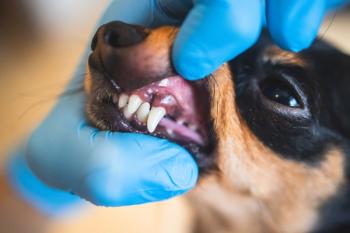
Crowded malaligned teeth and jaw size discrepancies are the most common contributors to malocclusion. Posterior crossbite occurs when the line of occlusion is incorrect buccolingually. Usually, the upper teeth are positioned lingually to the lower teeth. This is called lingual crossbite or just posterior crossbite. In buccal crossbite, which occurs rarely, if the upper teeth are too far buccally, there is no or little occlusal contact. This is a common finding in or line bred pocket pets and in some equine lines. The premolars and molars of some rodents can be seen to rotate 360 degrees extending into the orbit or turbinates in advance cases. Anterior cross bites are common in domestic canines. One or more upper incisors are displaced lingual to the lower incisors when the teeth are in occlusion...
A severe malocclusion may compromise all aspects of oral function. There may be difficulty in mastication if only a few teeth meet, and jaw discrepancies may force adaptive alterations in swallowing. Malocclusion contributes to both dental decay and periodontal disease. Plaque and food is retained in areas that are out of normal function making it harder to care for the teeth leading to pathology. Congenitally missing teeth. Congenital absence of teeth in dogs and cats can be the result of disturbances during the initial stages of formation of a tooth. Anodontia, the total absence of teeth, is the extreme form. The term oligodontia refers to congenital absence of many but not all teeth, whereas hypodontia implies the absence of only a few teeth. Since the primary teeth give rise to the permanent tooth buds, there will be no permanent tooth if its primary predecessor was missing. It is possible, however, for a primary tooth to be present and for the permanent counterpart to be absent. Anodontia or oligodontia in man is usually associated with an unusual but mild systemic abnormality, ectodermal dysplasia. Occasionally, oligodontia occurs in a patient with no apparent systemic problem or congenital syndrome.
Anodontia and oligodontia are rare, but hypodontia is a relatively common finding in dogs, especially pure bred and line bred dogs where the genetic fault has been perpetuated.. As a general rule, the premolar teeth are the most commonly missing, usually the second premolar. Rarely are the carnassial or canine teeth missing.
Malformed teeth are not as prevalent as supernumerary teeth. These abnormalities in tooth size or shape result from disturbances during the morph differentiation stage of development.
Supernumerary teeth also result from disturbances during the initiation and proliferation stages of dental development. Supernumery teeth may disrupt normal occlusal development and can contribute to periodontal disease. Supernumerary teeth, sclerotic bone, and heavy fibrous gingiva can also interfere with eruption. Extract only those supernumerary teeth that may contribute to malocclusion or crowding. If there is room for an extra tooth and its position does not present problems, it is not extracted. Dolicocephalic head types can accommodate extra premolars without crowding or occlusal disturbances.
Dental trauma can lead to the development of malocclusion. Damage to permanent tooth buds from an injury to primary teeth, drift of permanent teeth after premature loss of primary teeth, and direct injury to permanent teeth are primary causes. Trauma to a primary tooth can displace the developing permanent tooth bud or disrupt enamel formation with resultant coronal and or root defects. This root form is call dilaceration, defined as a distorted root form. Dilaceration can occur from any distortion of the crown relative to the root, and so may result from mechanical interference with eruption, but its usual cause, is trauma to primary teeth which also displaced the permanent buds.
It may be necessary to extract a severely dilacerated tooth.
A relationship between anatomic form and physiologic function is apparent in all animals. Over evolutionary time, adaptations in the jaws and dental apparatus are prominent in the fossil record.
Heredity versus developmental etiologic factors
Malocclusion is primarily the result of inherited dento-facial proportions governed by mans desire to breed the ideal dog or cat. These problems become established phenotypes with line and in breeding practices. They may be altered somewhat by developmental variations, trauma, or altered function.
Certain types of malocclusion are presented with certain breeds and head types. The prognathic mandible of the brachycephalic breeds for example. The pertinent question for the etiology of malocclusion is not whether there are in inherited influences on the jaws and teeth, because obviously there are, but whether malocclusion is often caused by inherited characteristics.
Malocclusion could be produced by inherited characteristics in two major possible ways. The first would be an inherited disproportion between the size of the teeth and the size of the jaws, which would produce crowding or spacing. The second possibility would be an inherited disproportion between size or shape of the upper and lower jaws, which would cause improper occlusal relationships. The more independently these characteristics are determined, the more likely that disproportions could be inherited. Large teeth but small jaws could be inherited for instance, or a large upper jaw and a small lower one. Conversely, if dento-facial characteristics tended to be linked, an inherited mismatch of this type would be unlikely.
Genetic isolation and uniformity as seen in the wild dogs produces rare instances of malocclusion. When all animals in a group carry the same genetic information for tooth size and jaw size, there would be no possibility of animals inheriting discordant characteristics. Genes that introduced disturbances into the masticatory system would tend to be eliminated from the population. The result should be exactly what is seen in breeds in whom tooth size-jaw size discrepancies are infrequent, and breeds in which everyone tends to have the same jaw relationship. When there is out breeding between originally distinct breeds malocclusal problems develop. This view of malocclusion as primarily a genetic problem was greatly strengthened by breeding experiments with animals carried out in the 1930s. By far the most influential individual in this regard was Professor Stockard, who methodically cross-bred dogs and recorded the interesting effects on body structure. Present-day dogs come in a variety of breeds and sizes.
Stockard felt that dramatic malocclusions did occur in cross-bred dogs, more from jaw discrepancies than from tooth size-jaw size imbalances. His research confirmed that "Independent inheritance of facial characteristics could be a major cause of malocclusion and that the rapid increase in malocclusion accompanying urbanization was probably the result of increased out breeding".
Stockards research did not take into account that many small breeds carry the gene for achondroplasia, a deficient growth of cartilage. The result may show extremely short legs and an underdeveloped midface. Most terriers and bulldogs also carry this gene. Achrondroplasia is an autosomal dominant trait; therefore the trait will be expressed more dramatically in some individuals than in others. Stockard explained that malocclusions were not based on inherited jaw size, but by the extent to which achondroplasia was expressed in that animal, and that severe malocclusions could be developed by crossing morphologically different breeds.
Modern orthodontic research has shown that the simple belief that malocclusion is the result of independent inheritance of dental and facial characteristics is not completely true. The precise role of heredity as an etiologic agent for malocclusion has not been clarified.
Certain conditions produce consistent occlusal problems. This includes, crowding of the teeth due to the continued breeding of animals with disproportionate in jaw and tooth size. Environmental factors play a role to some degree, however, and it is not clear what these are. There is no theoretical explanation of how a coarser diet and more powerful jaw function could significantly alter the dimensions of the dental arches. Open bite may be related to tongue posture, not to tongue activity during swallowing, as it is usually thrused against the upper teeth when swallowing
Anterior crossbite
The permanent incisors erupt lingual to the primary incisors. It was erroneously thought that prolonged retention of the primary incisors can deflect the permanent incisors lingually. Lingual displacement would arise by lingual tipping of the incisors as they erupt. Treatment is usually not necessary, unless there is periodontal involvement due to the crossbite.
Serial extraction hopes to correct a possible mechanical dental interlock of the incisors, or to intercept a developing malocclusion usually is not effective. The theory is that the removal or incisors prior to their normal time of exfoliation will allow the erupting permanent incisors to advance more rapidly foreword to catch up with the advanced counterpart. This procedure has limited merit. Also the extraction of primary canines however to intercept a developing problem has limited merit. True malocclusions cannot be intercepted by this procedure. The permanent lower canines erupt medial to the primary canines, while the permanent upper canines erupt anterior to the primary canines. It was thought that prolonged retention of the primary canines will force the permanent canines into malocclusion. The lower permanent canines will assume a base narrow position striking the palate upon closure, instead of occupying their normal position lateral to the maxilla, while the upper permanent canines will be forced into an anterior displacement. This in effect will prevent the lower canine form assuming its normal position.
Retention of primary teeth is a genetic fault. The erupting permanent replacement tooth is genetically malaligned. This presents as a base narrow position in the lower jaw. This lack of contact with the primary precursor tooth prevents root resorbtion. The result is both canines in the mouth at the same time
Extraction of the retained primary tooth is necessary. The permanent canine can then be moved into occlusion with orthodontic therapy.
Orthodontic movement of teeth can best be described as prolonged pressure application to a tooth Movement occurs as the bone around the tooth remodels. Bone is resorbed in areas of applied pressure, and remodeled at the opposite side of pressure. The tooth moves through the bone carrying its attachment apparatus with it, as the socket of the tooth migrates. Orthodontic movement requires resorbtion and apposition of bone adjacent to the root structure of teeth. Because the bony response is mediated by the periodontal ligament, tooth movement is primarily a function of the periodontal ligament. The gingival fiber networks are also disturbed by orthodontic tooth movement and must remodel to accommodate the new tooth positions. Elastic fibers occur in the gingiva, and reorganization occurs more slowly than the periodontal ligament itself. Collagenous fiber networks within the gingiva have normally completed their reorganization, but the elastic supracrestal fibers remodel much slower. Therefore it is possible for a tooth to recover part or all of its original position several months after relocation. These gingival fiber networks must remodel to accommodate the new tooth positions.
Orthodontic tooth movement is made possible by the application of prolonged forces. In addition, light prolonged forces in the natural environment, forces from the lips, cheeks, or tongue resting against the teeth, have the same potential as orthodontic forces to cause the teeth to move to a different location.
Light continuous force produces the most desired orthodontic tooth movement. This light force must be constant rather than intermittent. The behavior of orthodontic materials and mechanical factors must be considered in the design of orthodontic appliances that can deliver ideal light forces.
Bonding of orthodontic appliances to the teeth requires a mechanical locking of an adhesive to irregularities in the enamel surface of the tooth that is produces by acid etching techniques. Brackets can be locked to the teeth providing an attachment appliance for wires or elastics to be connected to a labial arch wire producing light directional forces. Careful attention to the components of the bonding system, acid etch techniques, bracket size and design, and tooth surface preparation, insure retention of bonded appliances.
Proper position for the attachment or bracket is predetermined and carried to place rapidly and accurately. The bonding cements usually set up in rather short working time.
References:
Stockard, C.R., and Johnson, A.L.: Genetic and Endocrine Basis for Differences in Form and Behavior. Philadelphia, 1941, the Wistar Institute of Anatomy and Biology, pp. 149-205.
Proffit,William R., Contemporary Orthodontics, Mosby,
Newsletter
From exam room tips to practice management insights, get trusted veterinary news delivered straight to your inbox—subscribe to dvm360.


