Otitis media and interna: Look for neurological signs
The practicing small animal veterinarian often has to face small animals with otitis externa. While not as common, otitis media and interna likely cause neurological signs.
The practicing small animal veterinarian often has to face small animals with otitis externa. While not as common, otitis media and interna likely cause neurological signs.
These signs may reflect peripheral or central nervous system involvement.
With otitis media, facial nerve and sympathetic nerve disturbances can be expected because these nerves pass through the wall of the middle ear and cavity of the middle ear, respectively (Veterinary Neuroanatomy and Clinical Neurology, Saunders 2nd ed., DeLahunta). Hence, in otitis media facial asymmetry, inability to blink, xerophthalmia and ipsilateral Horner's syndrome are the most likely neurological signs to accompany the head tilt. In otitis interna, one might observe balance problems (without conscious proprioceptive deficits), rotary or horizontal nystagmus with the fast phase away from the lesion (the lesion "slowly pulls" the eye toward the lesion side, and the fast phase is a "compensatory" phase, as the patient "jerks" the eye back to the normal position, toward the intact side). A key part of the neurological exam in the absence of spontaneous or resting nystagmus is to roll the patient on its back, extend the neck, and examine for the presence of positional nystagmus, which is also abnormal. Another possible neurological sign is the "eye drop" (ventrolateral strabismus) ipsilateral to the lesion (BSAVA Manual of Canine and Feline Neurology 3rd ed. SR Platt, NJ Olby). This abnormality, if present, can be elicited by raising the head. These neurological signs are most commonly associated with vestibulocochlear nerve involvement due to inner ear disease.
As otitis progresses, central nervous system involvement may occur. In a recent article (Clinical Signs, Magnetic Resonance Imaging Features, and Outcome After Surgical and Medical Treatment of Otogenic Intracranial Infection in 11 Cats and 4 Dogs, BK Sturges et al, J Vet Intern Med 2006; 20:648-656) the authors hypothesize that the major route of extension from the inner ear to the meninges and brain stem is through the internal acoustic meatus via blood vessels and nerves. Hematogeneous spread is also possible.
Once the infection involves the central nervous system, altered mental status (depression, stupor), seizures, cervical pain, paresis, proprioceptive deficits and ophistotonus may develop in addition to the previously discussed neurological signs.
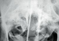
Neurological diagnostic tests following thorough neurological and physical examination include radiographs of the skull under general anesthesia (lateral, ventro dorsal or dorso ventral, oblique and open mouth). Photo 1 demonstrates increased soft-tissue density within the bulla on the right side of the radiograph in a patient with otitis interna. Other diagnostics indicated include otoscopic examination under general anesthesia (or video otoscopic exam), aerobic, anaerobic and cytological sample evaluation, cerebrospinal fluid analysis and magnetic resonance (MR) imaging. MR is the diagnostic imaging of choice if intracranial extension is suspected (BK Sturges, et al).

Photo 1 shows increased soft-tissue density within the bulla on the right side of the radiograph in a patient with otitis interna.
Besides inflammatory or infectious causes of otitis, tumors, foreign bodies (especially in the western U.S.) and idiopathic vestibular syndromes should be considered. It is noteworthy that in the article by Sturges et al, two out of three chronic otitis cases were diagnosed with cholesteatoma. Cholesteatomas are keratinizing masses in the tympanic cavity that may expand to the brain stem, and are believed secondary to chronic infections. Both dogs were treated surgically with bulla osteotomy, and the clinical signs resolved. But in one dog, the cholesteatoma recurred.
It is recommended that in patients with otitis media and interna refractory to conservative treatment (with or without central involvement) MR imaging and exploration of the bulla tympany should be considered in order to provide adequate drainage and to collect samples for histopathology and culture.
Dr. Nanai is a resident of the European College of Veterinary Neurology/Neurosurgery at the Animal Emergency and Referral Center in Fort Pierce, Fla.
Dr. Lyman is a graduate of The Ohio State University College of Veterinary Medicine. He completed a formal internship at the Animal Medical Center in New York City. Lyman is a co-author of chapters in the 2000 editions of Kirkâs Current Veterinary Therapy XIII and Quick Reference to Veterinary Medicine.