Parasites of red blood cells: Babesia and Mycoplasma (Proceedings)
Protozoal disease (genus: Babesia) of dogs and cats where merozoites (piroplasms) infect RBCs.
BABESIA
Overview
• Protozoal disease (genus: Babesia) of dogs and cats where merozoites (piroplasms) infect RBCs
• Degree of illness is usually dependent on the severity and rate of anemia development.
• Anemia mainly as a result of immune mediated hemolysis but also due to direct piroplasm damage to RBCs.
Etiology/Pathophysiology
• Infection (by tick transmission, transplacental, blood transfusion, or, in the case of B. gibsoni, by bite wounds presumably by blood from infected dog entering a bite wound.) followed by 2 week incubation period during which piroplasms infect and multiply in RBCs, resulting in RBC damage mainly from immune mediated processes but also direct RBC lysis.
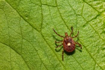
• In the case of Rhipicephalus sanguineus tick, larvae, adults and nymphs can all transmit infection; ticks need to be attached for several days; in ticks, the parasite reproduces by sexual reproduction; ticks can be infected by eating a blood meal or by transovarial means.
Dogs
• Large (4 – 7 µm): B. canis distributed worldwide; 3 sub-species based on biologic, genetic and geographic distribution:
o B. canisvogeli – USA, Africa, Asia, Australia. Transmitted by R. sanguineus ticks, so disease found in southeastern, southern states, and California.
o B. canis rossi - Africa (most virulent sub-species)
o B. canis canis – Europe, areas of Asia
o Small (2 – 5 µm): several genetically distinct sub-species:
o B. gibsoni – world-wide distribution (especially Asia) including the USA.
o B. conradae – (California – genetically distinct from B. gibsoni) – infects only dogs and only reported in California.
o B. microti-like – Spain, but recently reported in a Pit Bull terrier dog from Mississippi (unknown if this is a local case or imported).
o Theileria annae – (Spanish dog piroplasm). Reported in Spain and Europe.
Cats
• Small (2 – 5 µm) – B. felis reported in Africa.
Signalment/History
• History of tick attachment.
• History of recent dog bite wound may be a risk for B. gibsoni infection.
• Any age or breed of dog can be infected.
• Severity of disease – depends on the strain of the organism, and the age and breed of the animal.
• B. canis infections - more prevalent in Greyhounds (USA).
• B. gibsoni infections - more prevalent in American Pit Bull, Staffordshire, and Tosa Inu breeds.
Clinical Features - Dogs
• Peracute, acute, chronic, or asymptomatic (in some carrier animals).
• Splenectomy and immunosuppression – severely worsens disease (B. canis rossi) or makes it apparent (B. canis in the USA).
• Immunosuppression – may result in an increase in parasitemia and manifestation of clinical signs in chronically infected dogs (B. canis in the USA).
• Most severe disease – caused by B. gibsoni in the USA, and B. canis rossi in Africa.
• B. canis - rarely causes clinical disease in the USA.
Signs include: Lethargy, anorexia, weight loss, fever.
• Pale mucus membranes, icterus.
• Splenomegaly, lymphadenopathy, hemoglobinemia/uria.
• Gastrointestinal signs - Some dogs develop vomiting, diarrhea, dark feces (from increased bilirubin excretion).
• Cerebral babesiosis - weakness, disorientation, collapse (B. canis rossi in Africa).
• Renal/urologic disease – results in renal failure (B. canis rossi in Africa).
Differential Diagnosis
• Immune mediated diseases - hemolytic anemia, ITP, idiopathic, ehrlichiosis, RMSF, neoplasia, haemobartonellosis, cytauxzoonosis, SLE.
• Non-immune mediated diseases - hemolytic anemia; heartworm caval syndrome, zinc toxicity, splenic torsion, Heinz body anemia, DIC, PK deficiency, PFK deficiency.
• Causes of jaundice – hepatic or post-hepatic disease (obstruction or rupture of biliary tract).
Diagnostics
CBC
• Mild to severe (PCV < 10%) regenerative anemia.
• Peracute – animal may present before a regenerative response has time to occur.
• Anemia - may not be present in all cases (e.g. carriers: Greyhounds with B. canis in USA)
• Spherocytes, autoagglutination and positive Coombs test may also be present.
• Thrombocytopenia - usually moderate to severe, and can occur without anemia.
• Variable leukocytosis or leukopenia.
Biochemical profile/urinalysis
• Hyperbilirubinemia/uria – if hemolysis acute and severe (African cases rather than USA).
• Hyperglobulinemia - common in chronic cases (sometimes the only blood chemistry abnormality in these cases).
• Mild elevated liver enzymes - due to anemia/hypoxia.
• Renal failure and metabolic acidosis (B. canis rossi in Africa).
• Bilirubinuria – common.
• Hemoglobinuria – detected less commonly in the USA than in Africa.
Other tests
• Microscopic examination of stained thin or thick blood smears – can provide definitive diagnosis but sensitivity depends on experience of microscopist; modified Wright's stain best for viewing organism; blood from peripheral capillary (ear prick) may improve sensitivity; can not differentiate sub-species using microscopy.
• B. canis - large piriforms within RBCs but also ring forms.
• B. gibsoni - smaller and often single forms are found per RBC.
• Serology – IFA; false negatives in young dogs and in some infections; does not differentiate species and sub-species; use if microscopic examination negative with high clinical suspicion of disease, but if negative, need to confirm with a PCR.
• PCR - tests for presence of Babesia DNA in a biological sample (usually EDTA anti-coagulated whole blood), and can differentiate sub-species and species; more sensitive than microscopy.
• A combination of serology and PCR is considered to offer the highest sensitivity.
Therapeutics
• Anemic patients - transfusion of whole blood or packed RBCs (for loss of RBC mass).
• Polymerized bovine hemoglobin solution. May be used if fresh blood is not available.
• Severely affected patients require aggressive fluid therapy for hypovolemic shock from blood loss (usually as a result of thrombocytopenia with bleeding).
Drugs of choice: no drug regimen is 100% effective, so an infected dog is infected for life.
• Imidocarb diproprionate (Imizol® , Schering-Plough) - preferred therapy; may clear B. canis infections usually but not B. gibsoni (Asia). Certainly decrease morbidity and mortality
• Diminazine aceturate (not available in US and not FDA approved). Similar efficacy to Imazol®
• Metronidazole, clindamycin, and doxycycline: - decrease clinical signs but do not clear infection.
• Azithromycin – give in combination with atovaquone is preferred Tx for B. gibsoni infections.
• Prednisone - treat immune mediated component of anemia
ERYTHROCYTIC MYCOPLASMAL INFECTIONS
Definition/Overview
• Red blood cell destruction and anemia caused by parasite attachment to the external surface of RBCs and immune response by the host.
Etiology/Pathophysiology
• Haemobartonella felis (cats)and Haemobartonella canis (dogs) – classified at rickettsial bacteria.
• Recently recognized to be mycoplasmal bacteria based on genetic determinations.
• Proposed new names:-
o Mycoplasma haemofelis for a large form of H. felis.
o Mycoplasma haemominutum for the small form of H. felis.
o Mycoplasma turicensis – found on PCR only, not seen cytologically
o Mycoplasma haemocanis for H. canis (dogs).
• The large species of mycoplasmal organisms infecting cats – generally causes more severe disease than small species.
• Cats – anemia more severe if FeLV infected. Splenectomy does not make disease worse.
• Dogs – likelihood of severe anemia greatly increased if splenectomized or with pathologic changes in the spleen.
Signalment/History
• Worldwide distribtution.
• Most common in adult (dogs and cats).
• More common in males cats and FIV-infected cats.
• No sex prevalence in dogs.
Clinical Features
Cats
• Variable disease severity – ranges from inapparent infection to marked depression and death.
• Intermittent fever (50% of the time) during the acute phase.
• Depression, weakness, anorexia, pale mucous membranes.
• Splenomegaly.
• Icterus - rare.
Dogs
• Mild or inapparent signs – pale mucous membranes and listlessness.
• In splenectomized dogs – signs more like cats.
Differential Diagnosis
• Other causes of hemolytic anemia:- IMHA; babesiosis (not in cats in the U.S.); cytauxzoonosis (cats only); Heinz body hemolytic anemia; microangiopathic hemolytic anemia; pyruvate kinase deficiency; phosphofructokinase deficiency (dogs only).
• Differentiated from IMHA – only by recognition of parasites in blood (stained blood film or PCR-based assays); both disorders may be Coombs test-positive.
• Babesia and Cytauxzoon spp. – differ in morphology from these mycoplasmal organisms.
• New methylene blue stains – used to identify Heinz bodies.
• Enzyme assays or specialized DNA tests – used to diagnose pyruvate kinase and phosphofructokinase deficiencies.
Diagnostics
CBC
• Anemia – usually with reticulocytosis if regenerative. Also anisocytosis, macrocytosis, Howell-Jolly bodies, and occasionally marked normoblastemia.
• Anemia – may appear poorly regenerative if a precipitous decrease in PCV has occurred early in the disease or if there are other concurrent disorders (e.g., FeLV or FIV infections in cats).
• Autoagglutination – may see in feline blood samples after they cool to below body temperature.
• Variable total and differential leukocyte counts of little diagnostic assistance.
• Hemoglobinemia – rarely observed, so no hemoglobinuria reported.
Biochemical profile
• Hyperbilirubinemia – seen in some cases but seldom severe.
• Substantial bilirubinuria – seen in some dogs.
• Abnormalities related to anemic hypoxia or profile can be normal.
• Hypoglycemia – possible in moribund cats (not specific to this disease).
• Plasma protein concentrations – usually normal but may be increased.
• Routine blood stains (e.g., Wright - Giemsa) to identify organisms in blood films; overall, low sensitivity; examine before treatment is begun as treatment increases false negatives.
• Reticulocyte – stains can't be used as punctate reticulocytes (cats) appear similar to parasites.
• Organisms must be differentiated from precipitated stain – refractile drying or fixation artifacts, poorly staining Howell-Jolly bodies, and basophilic stippling can confuse diagnosis.
• Feline organisms – small blue-staining cocci, rings, or rods on RBCs.
• Canine organisms – commonly form chains of organisms that appear as filamentous structures on the surface of RBCs.
• Parasitemia – cyclic, and thus organisms not always identifiable in blood (especially in cats).
• PCR-based assays (on whole blood or dry) – significantly more sensitive than cytology.
• Direct Coombs test – may be positive.
Therapeutics
• Without therapy – mortality with the larger form may reach 30% in cats.
• Outpatient treatment – unless severely anemic or moribund.
• Blood transfusions – required when the anemia is considered life-threatening.
• IV administration of glucose-containing fluid – recommended in moribund animals.
Drugs of choice
• Doxycycline (10mg/kg/day, PO, for 2 wks) – drugs of choice.
• Enrofloxacin (5mg/kg/day, PO) – efficacious alternative to doxycycline.
• Prednisolone – may be given to severely anemic animals (to treat immune mediated destruction of RBCs); gradually decrease dosage as the PCV increases.
Precautions/interactions
• Tetracycline antibiotics – may produce fever or evidence of gastrointestinal disease in cats; use a lower dosage or a different drug, or discontinue drug therapy altogether.
• Doxycycline – may cause esophagitis and stricture in cats; use liquid form or give with food.
• Enrofloxacin – has been associated with blindness in cats; may cause arthropathy in young cats.
• Chloramphenicol – may use in dogs; can cause dose-dependent erythroid hypoplasia in cats.
Comments
• Examine animal after 1 week of treatment – to confirm that PCV has risen.
• Alert owner – cat may remain carriers even after completion of treatment but seldom relapse with disease once PCV returns to normal.
• Prognosis usually excellent in cats – if treated early in disease.
REFERENCES
1. Willi B, Boretti FS, Cattori V, et al: Identification, molecular characterization, and experimental transmission of a new hemoplasma isolate from a cat with hemolytic anemia in Switzerland. J Clin Microbiol 2005;43(6):2581-5.
2. Sykes JE, Drazenovich NL, Ball LM, et al: Use of conventional and real-time polymerase chain reaction to determine the epidemiology of hemoplasma infections in anemic and nonanemic cats. J Vet Intern Med 2007;21(4):685-93.
3. Tasker S, Peters IR, Papasouliotis K, et al. Description of outcomes of experimental infection with feline haemoplasmas: Copy numbers, haematology, Coombs' testing and blood glucose concentrations. Vet Microbiol 2009;139(3-4):323-332.
4. Sykes JE, Terry JC, Lindsay LL, et al: Prevalences of various hemoplasma species among cats in the United States with possible hemoplasmosis. J Am Vet Med Assoc 2008;232(3):372-9.
5. Jefferies R, Ryan UM, Jardine J, et al.Babesia gibsoni: Detection during experimental infections and after combined atovaquone and azithromycin therapy. Exp Parasitol 2007;117:115-23.
6. Irwin PJ. Canine babesiosis: from molecular taxonomy to control. Parasites & Vectors 2009;2 (S1):1-9.
Newsletter
From exam room tips to practice management insights, get trusted veterinary news delivered straight to your inbox—subscribe to dvm360.


